


 Foundational characteristics of cancer include proliferation, angiogenesis, migration, evasion of apoptosis, and cellular immortality. Find key markers for these cellular processes and antibodies to detect them.
 The SUMOplot™ Analysis Program predicts and scores sumoylation sites in your protein. SUMOylation is a post-translational modification involved in various cellular processes, such as nuclear-cytosolic transport, transcriptional regulation, apoptosis, protein stability, response to stress, and progression through the cell cycle.
 The Autophagy Receptor Motif Plotter predicts and scores autophagy receptor binding sites in your protein. Identifying proteins connected to this pathway is critical to understanding the role of autophagy in physiological as well as pathological processes such as development, differentiation, neurodegenerative diseases, stress, infection, and cancer.
Anti-NUDEL Rabbit Monoclonal Antibody
- SPECIFICATION
- CITATIONS
- PROTOCOLS
- BACKGROUND

Application
| WB, IHC |
|---|---|
| Primary Accession | Q9GZM8 |
| Host | Rabbit |
| Isotype | IgG |
| Reactivity | Rat, Human, Mouse |
| Clonality | Monoclonal |
| Format | Liquid |
| Description | Anti-NUDEL Rabbit Monoclonal Antibody . Tested in WB, IHC applications. This antibody reacts with Human, Mouse, Rat. |
| Gene ID | 81565 |
|---|---|
| Other Names | Nuclear distribution protein nudE-like 1, Protein Nudel, Mitosin-associated protein 1, NDEL1, EOPA, MITAP1, NUDEL |
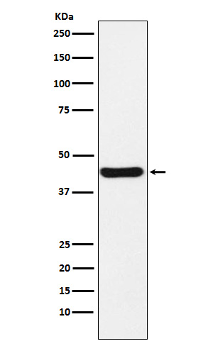
| Calculated MW | 40 kDa |
| Application Details | WB 1:500-1:2000 IHC 1:50-1:200 |
| Contents | Rabbit IgG in phosphate buffered saline, pH 7.4, 150mM NaCl, 0.02% sodium azide and 50% glycerol, 0.4-0.5mg/ml BSA. |
| Clone Names | Clone: 19N85 |
| Immunogen | A synthesized peptide derived from human NUDEL |
| Purification | Affinity-chromatography |
| Storage | Store at -20°C for one year. For short term storage and frequent use, store at 4°C for up to one month. Avoid repeated freeze-thaw cycles. |
| Name | NDEL1 |
|---|---|
| Synonyms | EOPA, MITAP1, NUDEL |
| Function | Required for organization of the cellular microtubule array and microtubule anchoring at the centrosome. May regulate microtubule organization at least in part by targeting the microtubule severing protein KATNA1 to the centrosome. Also positively regulates the activity of the minus-end directed microtubule motor protein dynein. May enhance dynein-mediated microtubule sliding by targeting dynein to the microtubule plus ends. Required for several dynein- and microtubule-dependent processes such as the maintenance of Golgi integrity, the centripetal motion of secretory vesicles and the coupling of the nucleus and centrosome. Also required during brain development for the migration of newly formed neurons from the ventricular/subventricular zone toward the cortical plate. Plays a role, together with DISC1, in the regulation of neurite outgrowth. Required for mitosis in some cell types but appears to be dispensible for mitosis in cortical neuronal progenitors, which instead requires NDE1. Facilitates the polymerization of neurofilaments from the individual subunits NEFH and NEFL. Positively regulates lysosome peripheral distribution and ruffled border formation in osteoclasts (By similarity). Plays a role, together with DISC1, in the regulation of neurite outgrowth (By similarity). May act as a RAB9A/B effector that tethers RAB9-associated late endosomes to the dynein motor for their retrograde transport to the trans-Golgi network (PubMed:34793709). |
| Cellular Location | Cytoplasm, cytoskeleton. Cytoplasm, cytoskeleton, microtubule organizing center, centrosome. Chromosome, centromere, kinetochore. Cytoplasm, cytoskeleton, spindle. Note=Localizes to the cell body of the motor neurons and colocalizes with assembled neurofilaments within axonal processes. Localizes to the microtubules of the manchette in elongated spermatids. Colocalizes with DISC1 in the perinuclear region, including the centrosome (By similarity). Localizes to the interphase centrosome and the mitotic spindle. Localizes to the kinetochore in a CENPF-dependent manner. |
| Tissue Location | Expressed in brain, heart, kidney, liver, lung, pancreas, placenta and skeletal muscle. |
Research Areas
Citations (0)

Thousands of laboratories across the world have published research that depended on the performance of antibodies from Abcepta to advance their research. Check out links to articles that cite our products in major peer-reviewed journals, organized by research category.
Submit your citation using an Abcepta antibody to
info@abcepta.com, and receive a free "I Love Antibodies" mug.
info@abcepta.com, and receive a free "I Love Antibodies" mug.

Application Protocols
Provided below are standard protocols that you may find useful for product applications.
Abcepta welcomes feedback from its customers.
If you have used an Abcepta product and would like to share how it has performed, please click on the "Submit Review" button and provide the requested information. Our staff will examine and post your review and contact you if needed.
If you have any additional inquiries please email technical services at tech@abcepta.com.
$ 370.00
Cat# ABO15409
Ordering Information
United States
AlbaniaAustraliaAustriaBelgiumBosnia & HerzegovinaBrazilBulgariaCanadaCentral AmericaChinaCroatiaCyprusCzech RepublicDenmarkEstoniaFinlandFranceGermanyGreeceHong KongHungaryIcelandIndiaIndonesiaIrelandIsraelItalyJapanLatviaLithuaniaLuxembourgMacedoniaMalaysiaMaltaMexicoNetherlandsNew ZealandNorwayPakistanPolandPortugalRomaniaSerbiaSingaporeSlovakiaSloveniaSouth AfricaSouth KoreaSpainSwedenSwitzerlandTaiwanTurkeyUnited KingdomUnited StatesVietnamWorldwideOthers
USA Headquarters
(888) 735-7227 / (858) 622-0099 or (858) 875-1900
Other Products
Shipping Information
Domestic orders (in stock items)
Shipped out the same day. Orders placed after 1 PM (PST) will ship out the next business day.
International orders
Contact your local distributors
