


 Foundational characteristics of cancer include proliferation, angiogenesis, migration, evasion of apoptosis, and cellular immortality. Find key markers for these cellular processes and antibodies to detect them.
 The SUMOplot™ Analysis Program predicts and scores sumoylation sites in your protein. SUMOylation is a post-translational modification involved in various cellular processes, such as nuclear-cytosolic transport, transcriptional regulation, apoptosis, protein stability, response to stress, and progression through the cell cycle.
 The Autophagy Receptor Motif Plotter predicts and scores autophagy receptor binding sites in your protein. Identifying proteins connected to this pathway is critical to understanding the role of autophagy in physiological as well as pathological processes such as development, differentiation, neurodegenerative diseases, stress, infection, and cancer.
> home > Products > Primary Antibodies > Signal Transduction > Anti-HIPK2 Rabbit Monoclonal Antibody
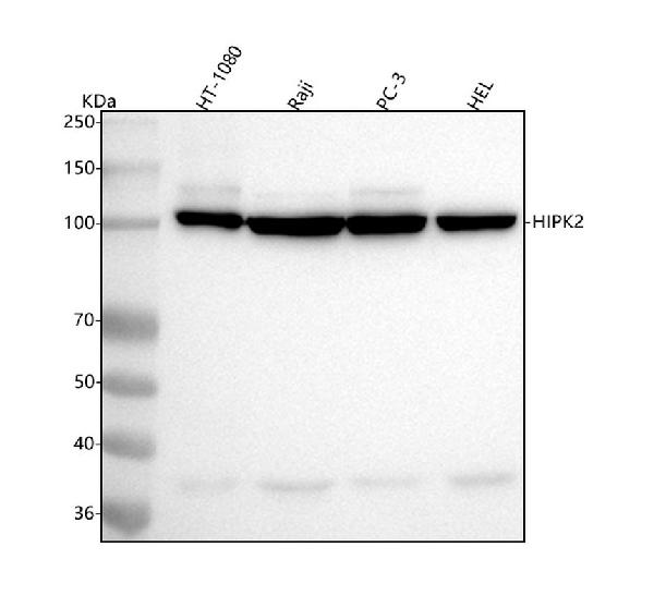
Anti-HIPK2 Rabbit Monoclonal Antibody
- SPECIFICATION
- CITATIONS
- PROTOCOLS
- BACKGROUND

Application
| WB, IF, ICC, IP, FC |
|---|---|
| Primary Accession | Q9H2X6 |
| Host | Rabbit |
| Isotype | IgG |
| Reactivity | Human, Mouse |
| Clonality | Monoclonal |
| Format | Liquid |
| Description | Anti-HIPK2 Rabbit Monoclonal Antibody . Tested in WB, ICC/IF, IP, Flow Cytometry applications. This antibody reacts with Human, Mouse. |
| Gene ID | 28996 |
|---|---|
| Other Names | Homeodomain-interacting protein kinase 2, hHIPk2, 2.7.11.1, HIPK2 |
| Calculated MW | 130 kDa, 100 kDa |
| Application Details | WB 1:500-1:2000 ICC/IF 1:50-1:200 IP 1:30 FC 1:500 |
| Contents | Rabbit IgG in phosphate buffered saline, pH 7.4, 150mM NaCl, 0.02% sodium azide and 50% glycerol, 0.4-0.5mg/ml BSA. |
| Clone Names | Clone: 21H33 |
| Immunogen | A synthesized peptide derived from human HIPK2 |
| Purification | Affinity-chromatography |
| Storage | Store at -20°C for one year. For short term storage and frequent use, store at 4°C for up to one month. Avoid repeated freeze-thaw cycles. |
| Name | HIPK2 |
|---|---|
| Function | Serine/threonine-protein kinase involved in transcription regulation, p53/TP53-mediated cellular apoptosis and regulation of the cell cycle. Acts as a corepressor of several transcription factors, including SMAD1 and POU4F1/Brn3a and probably NK homeodomain transcription factors. Phosphorylates PDX1, ATF1, PML, p53/TP53, CREB1, CTBP1, CBX4, RUNX1, EP300, CTNNB1, HMGA1, ZBTB4 and DAZAP2. Inhibits cell growth and promotes apoptosis through the activation of p53/TP53 both at the transcription level and at the protein level (by phosphorylation and indirect acetylation). The phosphorylation of p53/TP53 may be mediated by a p53/TP53-HIPK2-AXIN1 complex. Involved in the response to hypoxia by acting as a transcriptional co-suppressor of HIF1A. Mediates transcriptional activation of TP73. In response to TGFB, cooperates with DAXX to activate JNK. Negative regulator through phosphorylation and subsequent proteasomal degradation of CTNNB1 and the antiapoptotic factor CTBP1. In the Wnt/beta-catenin signaling pathway acts as an intermediate kinase between MAP3K7/TAK1 and NLK to promote the proteasomal degradation of MYB. Phosphorylates CBX4 upon DNA damage and promotes its E3 SUMO-protein ligase activity. Activates CREB1 and ATF1 transcription factors by phosphorylation in response to genotoxic stress. In response to DNA damage, stabilizes PML by phosphorylation. PML, HIPK2 and FBXO3 may act synergically to activate p53/TP53-dependent transactivation. Promotes angiogenesis, and is involved in erythroid differentiation, especially during fetal liver erythropoiesis. Phosphorylation of RUNX1 and EP300 stimulates EP300 transcription regulation activity. Triggers ZBTB4 protein degradation in response to DNA damage. In response to DNA damage, phosphorylates DAZAP2 which localizes DAZAP2 to the nucleus, reduces interaction of DAZAP2 with HIPK2 and prevents DAZAP2-dependent ubiquitination of HIPK2 by E3 ubiquitin-protein ligase SIAH1 and subsequent proteasomal degradation (PubMed:33591310). Modulates HMGA1 DNA-binding affinity. In response to high glucose, triggers phosphorylation-mediated subnuclear localization shifting of PDX1. Involved in the regulation of eye size, lens formation and retinal lamination during late embryogenesis. |
| Cellular Location | Nucleus, PML body. Cytoplasm Cytoplasm, Stress granule Note=Concentrated in PML/POD/ND10 nuclear bodies. Small amounts are cytoplasmic |
| Tissue Location | Highly expressed in heart, muscle and kidney. Weakly expressed in a ubiquitous way. Down-regulated in several thyroid and breast tumors. |
Research Areas
Citations (0)

Thousands of laboratories across the world have published research that depended on the performance of antibodies from Abcepta to advance their research. Check out links to articles that cite our products in major peer-reviewed journals, organized by research category.
Submit your citation using an Abcepta antibody to
info@abcepta.com, and receive a free "I Love Antibodies" mug.
info@abcepta.com, and receive a free "I Love Antibodies" mug.

Application Protocols
Provided below are standard protocols that you may find useful for product applications.
Abcepta welcomes feedback from its customers.
If you have used an Abcepta product and would like to share how it has performed, please click on the "Submit Review" button and provide the requested information. Our staff will examine and post your review and contact you if needed.
If you have any additional inquiries please email technical services at tech@abcepta.com.
$ 370.00
Cat# ABO15557
Ordering Information
United States
AlbaniaAustraliaAustriaBelgiumBosnia & HerzegovinaBrazilBulgariaCanadaCentral AmericaChinaCroatiaCyprusCzech RepublicDenmarkEstoniaFinlandFranceGermanyGreeceHong KongHungaryIcelandIndiaIndonesiaIrelandIsraelItalyJapanLatviaLithuaniaLuxembourgMacedoniaMalaysiaMaltaMexicoNetherlandsNew ZealandNorwayPakistanPolandPortugalRomaniaSerbiaSingaporeSlovakiaSloveniaSouth AfricaSouth KoreaSpainSwedenSwitzerlandTaiwanTurkeyUnited KingdomUnited StatesVietnamWorldwideOthers
USA Headquarters
(888) 735-7227 / (858) 622-0099 or (858) 875-1900
Other Products
Shipping Information
Domestic orders (in stock items)
Shipped out the same day. Orders placed after 1 PM (PST) will ship out the next business day.
International orders
Contact your local distributors
