


 Foundational characteristics of cancer include proliferation, angiogenesis, migration, evasion of apoptosis, and cellular immortality. Find key markers for these cellular processes and antibodies to detect them.
 The SUMOplot™ Analysis Program predicts and scores sumoylation sites in your protein. SUMOylation is a post-translational modification involved in various cellular processes, such as nuclear-cytosolic transport, transcriptional regulation, apoptosis, protein stability, response to stress, and progression through the cell cycle.
 The Autophagy Receptor Motif Plotter predicts and scores autophagy receptor binding sites in your protein. Identifying proteins connected to this pathway is critical to understanding the role of autophagy in physiological as well as pathological processes such as development, differentiation, neurodegenerative diseases, stress, infection, and cancer.
> home > Products > Primary Antibodies > Antibody Collections > SARS-Human interactions > Anti-TMPRSS2 Rabbit Monoclonal Antibody
Anti-TMPRSS2 Rabbit Monoclonal Antibody
- SPECIFICATION
- CITATIONS
- PROTOCOLS
- BACKGROUND

Application
| WB, IHC, IF, ICC, FC |
|---|---|
| Primary Accession | O15393 |
| Host | Rabbit |
| Isotype | IgG |
| Reactivity | Rat, Human, Mouse |
| Clonality | Monoclonal |
| Format | Liquid |
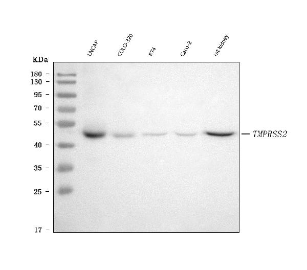
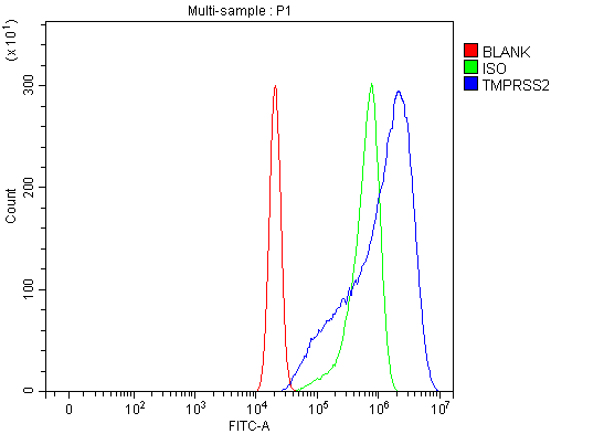
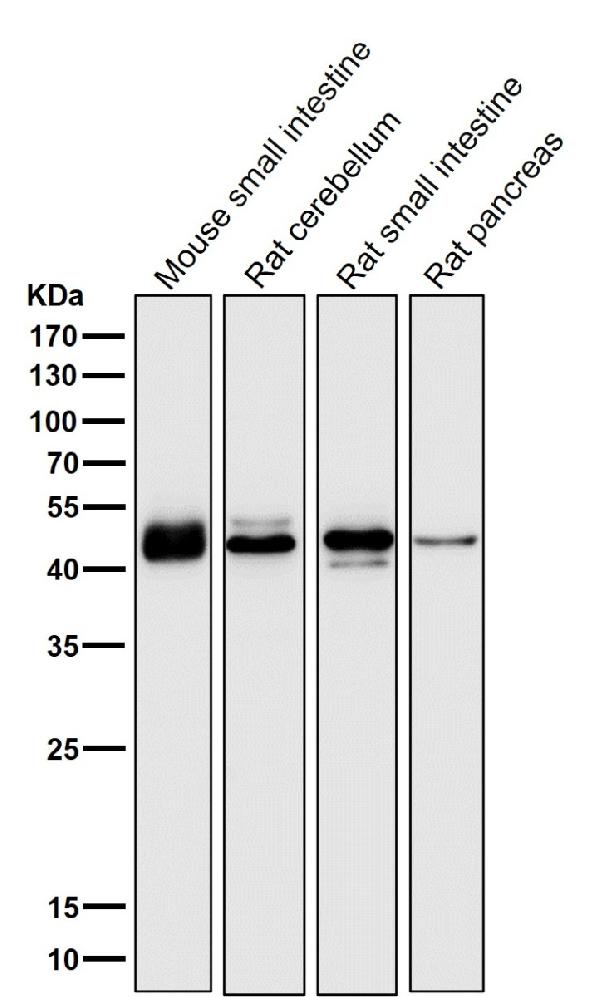
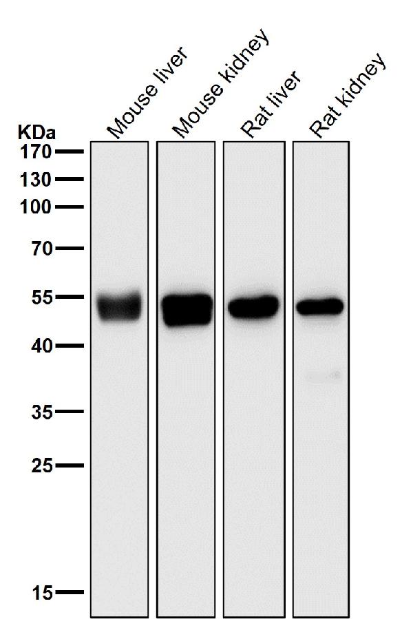
| Description | Anti-TMPRSS2 Rabbit Monoclonal Antibody . Tested in WB, IHC, ICC/IF, Flow Cytometry applications. This antibody reacts with Human, Mouse, Rat. |
| Gene ID | 7113 |
|---|---|
| Other Names | Transmembrane protease serine 2, 3.4.21.122, Serine protease 10, Transmembrane protease serine 2 non-catalytic chain, Transmembrane protease serine 2 catalytic chain, TMPRSS2 (HGNC:11876), PRSS10 |
| Calculated MW | 54 kDa |
| Application Details | WB 1:500-1:2000 IHC 1:100-1:500 ICC/IF 1:100-1:500 FC 1:60 |
| Contents | Rabbit IgG in phosphate buffered saline, pH 7.4, 150mM NaCl, 0.02% sodium azide and 50% glycerol, 0.4-0.5mg/ml BSA. |
| Clone Names | Clone: 23T34 |
| Immunogen | A synthesized peptide derived from human TMPRSS2 |
| Purification | Affinity-chromatography |
| Storage | Store at -20°C for one year. For short term storage and frequent use, store at 4°C for up to one month. Avoid repeated freeze-thaw cycles. |
| Name | TMPRSS2 (HGNC:11876) |
|---|---|
| Synonyms | PRSS10 |
| Function | Plasma membrane-anchored serine protease that cleaves at arginine residues (PubMed:32703818, PubMed:35676539, PubMed:37990007, PubMed:38964328). Participates in proteolytic cascades of relevance for the normal physiologic function of the prostate (PubMed:25122198). Androgen-induced TMPRSS2 activates several substrates that include pro- hepatocyte growth factor/HGF, the protease activated receptor-2/F2RL1 or matriptase/ST14 leading to extracellular matrix disruption and metastasis of prostate cancer cells (PubMed:15537383, PubMed:25122198, PubMed:26018085). In addition, activates trigeminal neurons and contribute to both spontaneous pain and mechanical allodynia (By similarity). |
| Cellular Location | Cell membrane; Single-pass type II membrane protein |
| Tissue Location | Expressed in several tissues that comprise large populations of epithelial cells with the highest level of transcripts measured in the prostate gland. Expressed in type II pneumocytes in the lung (at protein level). Expressed strongly in small intestine. Also expressed in colon, stomach and salivary gland. Coexpressed with ACE2 within lung type II pneumocytes, ileal absorptive enterocytes, intestinal epithelial cells, cornea, gallbladder and nasal goblet secretory cells (Ref.21). {ECO:0000269|PubMed:11169526, ECO:0000269|PubMed:20382709, ECO:0000269|PubMed:21325420, ECO:0000269|PubMed:32404436, ECO:0000269|Ref.21} |
Research Areas
Citations (0)

Thousands of laboratories across the world have published research that depended on the performance of antibodies from Abcepta to advance their research. Check out links to articles that cite our products in major peer-reviewed journals, organized by research category.
Submit your citation using an Abcepta antibody to
info@abcepta.com, and receive a free "I Love Antibodies" mug.
info@abcepta.com, and receive a free "I Love Antibodies" mug.

Application Protocols
Provided below are standard protocols that you may find useful for product applications.
Abcepta welcomes feedback from its customers.
If you have used an Abcepta product and would like to share how it has performed, please click on the "Submit Review" button and provide the requested information. Our staff will examine and post your review and contact you if needed.
If you have any additional inquiries please email technical services at tech@abcepta.com.
$ 370.00
Cat# ABO15758
Ordering Information
United States
AlbaniaAustraliaAustriaBelgiumBosnia & HerzegovinaBrazilBulgariaCanadaCentral AmericaChinaCroatiaCyprusCzech RepublicDenmarkEstoniaFinlandFranceGermanyGreeceHong KongHungaryIcelandIndiaIndonesiaIrelandIsraelItalyJapanLatviaLithuaniaLuxembourgMacedoniaMalaysiaMaltaMexicoNetherlandsNew ZealandNorwayPakistanPolandPortugalRomaniaSerbiaSingaporeSlovakiaSloveniaSouth AfricaSouth KoreaSpainSwedenSwitzerlandTaiwanTurkeyUnited KingdomUnited StatesVietnamWorldwideOthers
USA Headquarters
(888) 735-7227 / (858) 622-0099 or (858) 875-1900
Other Products
Shipping Information
Domestic orders (in stock items)
Shipped out the same day. Orders placed after 1 PM (PST) will ship out the next business day.
International orders
Contact your local distributors
