


 Foundational characteristics of cancer include proliferation, angiogenesis, migration, evasion of apoptosis, and cellular immortality. Find key markers for these cellular processes and antibodies to detect them.
 The SUMOplot™ Analysis Program predicts and scores sumoylation sites in your protein. SUMOylation is a post-translational modification involved in various cellular processes, such as nuclear-cytosolic transport, transcriptional regulation, apoptosis, protein stability, response to stress, and progression through the cell cycle.
 The Autophagy Receptor Motif Plotter predicts and scores autophagy receptor binding sites in your protein. Identifying proteins connected to this pathway is critical to understanding the role of autophagy in physiological as well as pathological processes such as development, differentiation, neurodegenerative diseases, stress, infection, and cancer.
Anti-ESE1 Rabbit Monoclonal Antibody
- SPECIFICATION
- CITATIONS
- PROTOCOLS
- BACKGROUND

Application
| WB, FC |
|---|---|
| Primary Accession | P78545 |
| Host | Rabbit |
| Isotype | IgG |
| Reactivity | Rat, Human, Mouse |
| Clonality | Monoclonal |
| Format | Liquid |
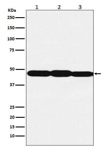
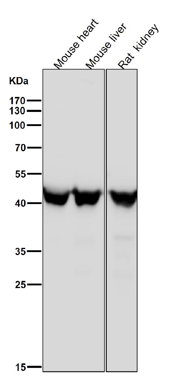
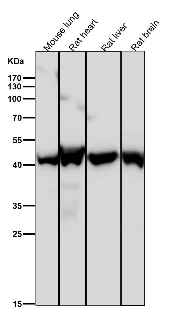
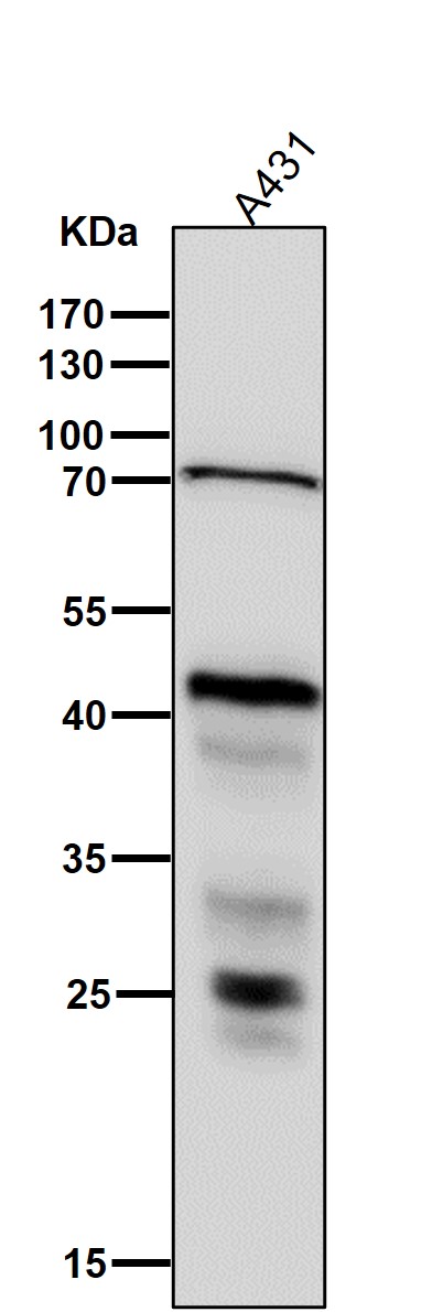
| Description | Anti-ESE1 Rabbit Monoclonal Antibody . Tested in WB, Flow Cytometry applications. This antibody reacts with Human, Mouse, Rat. |
| Gene ID | 1999 |
|---|---|
| Other Names | ETS-related transcription factor Elf-3, E74-like factor 3, Epithelial-restricted with serine box, Epithelium-restricted Ets protein ESX, Epithelium-specific Ets transcription factor 1, ESE-1, ELF3 (HGNC:3318) |
| Calculated MW | 47 kDa |
| Application Details | WB 1:500-1:2000 FC 1:100 |
| Contents | Rabbit IgG in phosphate buffered saline, pH 7.4, 150mM NaCl, 0.02% sodium azide and 50% glycerol, 0.4-0.5mg/ml BSA. |
| Clone Names | Clone: 24E94 |
| Immunogen | A synthesized peptide derived from human ESE1 |
| Purification | Affinity-chromatography |
| Storage | Store at -20°C for one year. For short term storage and frequent use, store at 4°C for up to one month. Avoid repeated freeze-thaw cycles. |
| Name | ELF3 (HGNC:3318) |
|---|---|
| Function | Transcriptional activator that binds and transactivates ETS sequences containing the consensus nucleotide core sequence GGA[AT]. Acts synergistically with POU2F3 to transactivate the SPRR2A promoter and with RUNX1 to transactivate the ANGPT1 promoter. Also transactivates collagenase, CCL20, CLND7, FLG, KRT8, NOS2, PTGS2, SPRR2B, TGFBR2 and TGM3 promoters. Represses KRT4 promoter activity. Involved in mediating vascular inflammation. May play an important role in epithelial cell differentiation and tumorigenesis. May be a critical downstream effector of the ERBB2 signaling pathway. May be associated with mammary gland development and involution. Plays an important role in the regulation of transcription with TATA-less promoters in preimplantation embryos, which is essential in preimplantation development (By similarity). |
| Cellular Location | Cytoplasm. Nucleus {ECO:0000255|PROSITE-ProRule:PRU00237, ECO:0000269|PubMed:10391676, ECO:0000269|PubMed:15169914, ECO:0000269|PubMed:17060315} Note=Localizes to the cytoplasm where it has been shown to transform MCF-12A mammary epithelial cells via a novel cytoplasmic mechanism Also transiently expressed and localized to the nucleus where it induces apoptosis in non-transformed breast epithelial cells MCF-10A and MCF-12A via a transcription-dependent mechanism |
| Tissue Location | Expressed exclusively in tissues containing a high content of terminally differentiated epithelial cells including mammary gland, colon, trachea, kidney, prostate, uterus, stomach and skin |
Citations (0)

Thousands of laboratories across the world have published research that depended on the performance of antibodies from Abcepta to advance their research. Check out links to articles that cite our products in major peer-reviewed journals, organized by research category.
Submit your citation using an Abcepta antibody to
info@abcepta.com, and receive a free "I Love Antibodies" mug.
info@abcepta.com, and receive a free "I Love Antibodies" mug.

Application Protocols
Provided below are standard protocols that you may find useful for product applications.
Abcepta welcomes feedback from its customers.
If you have used an Abcepta product and would like to share how it has performed, please click on the "Submit Review" button and provide the requested information. Our staff will examine and post your review and contact you if needed.
If you have any additional inquiries please email technical services at tech@abcepta.com.
$ 370.00
Cat# ABO15917
Ordering Information
United States
AlbaniaAustraliaAustriaBelgiumBosnia & HerzegovinaBrazilBulgariaCanadaCentral AmericaChinaCroatiaCyprusCzech RepublicDenmarkEstoniaFinlandFranceGermanyGreeceHong KongHungaryIcelandIndiaIndonesiaIrelandIsraelItalyJapanLatviaLithuaniaLuxembourgMacedoniaMalaysiaMaltaMexicoNetherlandsNew ZealandNorwayPakistanPolandPortugalRomaniaSerbiaSingaporeSlovakiaSloveniaSouth AfricaSouth KoreaSpainSwedenSwitzerlandTaiwanTurkeyUnited KingdomUnited StatesVietnamWorldwideOthers
USA Headquarters
(888) 735-7227 / (858) 622-0099 or (858) 875-1900
Other Products
Shipping Information
Domestic orders (in stock items)
Shipped out the same day. Orders placed after 1 PM (PST) will ship out the next business day.
International orders
Contact your local distributors
