


 Foundational characteristics of cancer include proliferation, angiogenesis, migration, evasion of apoptosis, and cellular immortality. Find key markers for these cellular processes and antibodies to detect them.
 The SUMOplot™ Analysis Program predicts and scores sumoylation sites in your protein. SUMOylation is a post-translational modification involved in various cellular processes, such as nuclear-cytosolic transport, transcriptional regulation, apoptosis, protein stability, response to stress, and progression through the cell cycle.
 The Autophagy Receptor Motif Plotter predicts and scores autophagy receptor binding sites in your protein. Identifying proteins connected to this pathway is critical to understanding the role of autophagy in physiological as well as pathological processes such as development, differentiation, neurodegenerative diseases, stress, infection, and cancer.
> home > Products > Primary Antibodies > Signal Transduction > Anti-RGAP1 Rabbit Monoclonal Antibody
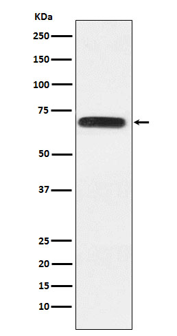
Anti-RGAP1 Rabbit Monoclonal Antibody
- SPECIFICATION
- CITATIONS
- PROTOCOLS
- BACKGROUND

Application
| WB, IHC, IF, ICC |
|---|---|
| Primary Accession | Q9H0H5 |
| Host | Rabbit |
| Isotype | IgG |
| Reactivity | Human |
| Clonality | Monoclonal |
| Format | Liquid |
| Description | Anti-RGAP1 Rabbit Monoclonal Antibody . Tested in WB, IHC, ICC/IF applications. This antibody reacts with Human. |
| Gene ID | 29127 |
|---|---|
| Other Names | Rac GTPase-activating protein 1, Male germ cell RacGap, MgcRacGAP, Protein CYK4 homolog, CYK4, HsCYK-4, RACGAP1 (HGNC:9804) |
| Calculated MW | 71 kDa |
| Application Details | WB 1:500-1:2000 IHC 1:50-1:200 ICC/IF 1:50-1:200 |
| Contents | Rabbit IgG in phosphate buffered saline, pH 7.4, 150mM NaCl, 0.02% sodium azide and 50% glycerol, 0.4-0.5mg/ml BSA. |
| Clone Names | Clone: 25R53 |
| Immunogen | A synthesized peptide derived from human RGAP1 |
| Purification | Affinity-chromatography |
| Storage | Store at -20°C for one year. For short term storage and frequent use, store at 4°C for up to one month. Avoid repeated freeze-thaw cycles. |
| Name | RACGAP1 (HGNC:9804) |
|---|---|
| Function | Component of the centralspindlin complex that serves as a microtubule-dependent and Rho-mediated signaling required for the myosin contractile ring formation during the cell cycle cytokinesis. Required for proper attachment of the midbody to the cell membrane during cytokinesis. Sequentially binds to ECT2 and RAB11FIP3 which regulates cleavage furrow ingression and abscission during cytokinesis (PubMed:18511905). Plays key roles in controlling cell growth and differentiation of hematopoietic cells through mechanisms other than regulating Rac GTPase activity (PubMed:10979956). Has a critical role in erythropoiesis (PubMed:34818416). Also involved in the regulation of growth-related processes in adipocytes and myoblasts. May be involved in regulating spermatogenesis and in the RACGAP1 pathway in neuronal proliferation. Shows strong GAP (GTPase activation) activity towards CDC42 and RAC1 and less towards RHOA. Essential for the early stages of embryogenesis. May play a role in regulating cortical activity through RHOA during cytokinesis. May participate in the regulation of sulfate transport in male germ cells. |
| Cellular Location | Nucleus. Cytoplasm. Cytoplasm, cytoskeleton, spindle Cytoplasmic vesicle, secretory vesicle, acrosome. Cleavage furrow Midbody, Midbody ring. Cell membrane; Peripheral membrane protein; Cytoplasmic side. Note=Colocalizes with RND2 in Golgi-derived proacrosomal vesicles and the acrosome (By similarity). During interphase, localized to the nucleus and cytoplasm along with microtubules, in anaphase, is redistributed to the central spindle and, in telophase and cytokinesis, to the midbody ring, also called Flemming body. Colocalizes with RHOA at the myosin contractile ring during cytokinesis. Colocalizes with ECT2 to the mitotic spindles during anaphase/metaphase, the cleavage furrow during telophase and at the midbody at the end of cytokinesis. Colocalizes with Cdc42 to spindle microtubules from prometaphase to telophase. |
| Tissue Location | Highly expressed in testis, thymus and placenta. Expressed at lower levels in spleen and peripheral blood lymphocytes In testis, expression is restricted to germ cells with the highest levels of expression found in spermatocytes. Expression is regulated in a cell cycle-dependent manner and peaks during G2/M phase |
Research Areas
Citations (0)

Thousands of laboratories across the world have published research that depended on the performance of antibodies from Abcepta to advance their research. Check out links to articles that cite our products in major peer-reviewed journals, organized by research category.
Submit your citation using an Abcepta antibody to
info@abcepta.com, and receive a free "I Love Antibodies" mug.
info@abcepta.com, and receive a free "I Love Antibodies" mug.

Application Protocols
Provided below are standard protocols that you may find useful for product applications.
Abcepta welcomes feedback from its customers.
If you have used an Abcepta product and would like to share how it has performed, please click on the "Submit Review" button and provide the requested information. Our staff will examine and post your review and contact you if needed.
If you have any additional inquiries please email technical services at tech@abcepta.com.
$ 370.00
Cat# ABO15974
Ordering Information
United States
AlbaniaAustraliaAustriaBelgiumBosnia & HerzegovinaBrazilBulgariaCanadaCentral AmericaChinaCroatiaCyprusCzech RepublicDenmarkEstoniaFinlandFranceGermanyGreeceHong KongHungaryIcelandIndiaIndonesiaIrelandIsraelItalyJapanLatviaLithuaniaLuxembourgMacedoniaMalaysiaMaltaMexicoNetherlandsNew ZealandNorwayPakistanPolandPortugalRomaniaSerbiaSingaporeSlovakiaSloveniaSouth AfricaSouth KoreaSpainSwedenSwitzerlandTaiwanTurkeyUnited KingdomUnited StatesVietnamWorldwideOthers
USA Headquarters
(888) 735-7227 / (858) 622-0099 or (858) 875-1900
Other Products
Shipping Information
Domestic orders (in stock items)
Shipped out the same day. Orders placed after 1 PM (PST) will ship out the next business day.
International orders
Contact your local distributors
