


 Foundational characteristics of cancer include proliferation, angiogenesis, migration, evasion of apoptosis, and cellular immortality. Find key markers for these cellular processes and antibodies to detect them.
 The SUMOplot™ Analysis Program predicts and scores sumoylation sites in your protein. SUMOylation is a post-translational modification involved in various cellular processes, such as nuclear-cytosolic transport, transcriptional regulation, apoptosis, protein stability, response to stress, and progression through the cell cycle.
 The Autophagy Receptor Motif Plotter predicts and scores autophagy receptor binding sites in your protein. Identifying proteins connected to this pathway is critical to understanding the role of autophagy in physiological as well as pathological processes such as development, differentiation, neurodegenerative diseases, stress, infection, and cancer.
> home > Products > Primary Antibodies > Signal Transduction > Anti-PLA2G2A Rabbit Monoclonal Antibody
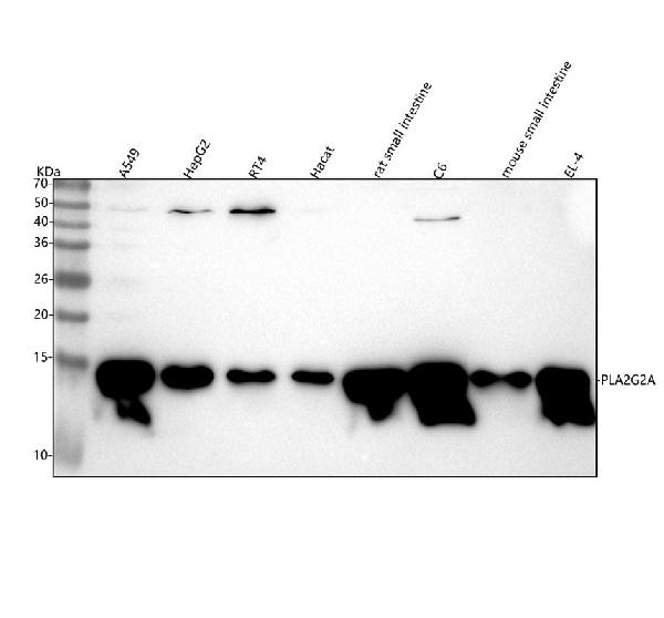
Anti-PLA2G2A Rabbit Monoclonal Antibody
- SPECIFICATION
- CITATIONS
- PROTOCOLS
- BACKGROUND

Application
| WB |
|---|---|
| Primary Accession | P14555 |
| Host | Rabbit |
| Isotype | IgG |
| Reactivity | Rat, Human, Mouse |
| Clonality | Monoclonal |
| Format | Liquid |
| Description | Anti-PLA2G2A Rabbit Monoclonal Antibody . Tested in WB application. This antibody reacts with Human, Mouse, Rat. |
| Gene ID | 5320 |
|---|---|
| Other Names | Phospholipase A2, membrane associated, 3.1.1.4, GIIC sPLA2, Group IIA phospholipase A2, Non-pancreatic secretory phospholipase A2, NPS-PLA2, Phosphatidylcholine 2-acylhydrolase 2A, PLA2G2A, PLA2B, PLA2L, RASF-A |
| Calculated MW | 14 kDa |
| Application Details | WB 1:500-1:2000 |
| Contents | Rabbit IgG in phosphate buffered saline, pH 7.4, 150mM NaCl, 0.02% sodium azide and 50% glycerol, 0.4-0.5mg/ml BSA. |
| Clone Names | Clone: 25P80 |
| Immunogen | A synthesized peptide derived from human PLA2G2A |
| Purification | Affinity-chromatography |
| Storage | Store at -20°C for one year. For short term storage and frequent use, store at 4°C for up to one month. Avoid repeated freeze-thaw cycles. |
| Name | PLA2G2A |
|---|---|
| Synonyms | PLA2B, PLA2L, RASF-A |
| Function | Secretory calcium-dependent phospholipase A2 that primarily targets extracellular phospholipids with implications in host antimicrobial defense, inflammatory response and tissue regeneration (PubMed:10455175, PubMed:10681567, PubMed:2925633). Hydrolyzes the ester bond of the fatty acyl group attached at sn-2 position of phospholipids (phospholipase A2 activity) with preference for phosphatidylethanolamines and phosphatidylglycerols over phosphatidylcholines (PubMed:10455175, PubMed:10681567). Contributes to lipid remodeling of cellular membranes and generation of lipid mediators involved in pathogen clearance. Displays bactericidal activity against Gram-positive bacteria by directly hydrolyzing phospholipids of the bacterial membrane (PubMed:10358193, PubMed:11694541). Upon sterile inflammation, targets membrane phospholipids of extracellular mitochondria released from activated platelets, generating free unsaturated fatty acids such as arachidonate that is used by neighboring leukocytes to synthesize inflammatory eicosanoids such as leukotrienes. Simultaneously, by compromising mitochondrial membrane integrity, promotes the release in circulation of potent damage-associated molecular pattern molecules that activate the innate immune response (PubMed:25082876). Plays a stem cell regulator role in the intestinal crypt. Within intracellular compartment mediates Paneth cell differentiation and its stem cell supporting functions by inhibiting Wnt signaling pathway in intestinal stem cell (ICS). Secreted in the intestinal lumen upon inflammation, acts in an autocrine way and promotes prostaglandin E2 synthesis that stimulates Wnt signaling pathway in ICS cells and tissue regeneration (By similarity). May play a role in the biosynthesis of N-acyl ethanolamines that regulate energy metabolism and inflammation. Hydrolyzes N-acyl phosphatidylethanolamines to N-acyl lysophosphatidylethanolamines, which are further cleaved by a lysophospholipase D to release N-acyl ethanolamines (PubMed:14998370). Independent of its catalytic activity, acts as a ligand for integrins (PubMed:18635536, PubMed:25398877). Binds to and activates integrins ITGAV:ITGB3, ITGA4:ITGB1 and ITGA5:ITGB1 (PubMed:18635536, PubMed:25398877). Binds to a site (site 2) which is distinct from the classical ligand-binding site (site 1) and induces integrin conformational changes and enhanced ligand binding to site 1 (PubMed:25398877). Induces cell proliferation in an integrin-dependent manner (PubMed:18635536). |
| Cellular Location | Secreted. Cell membrane; Peripheral membrane protein. Mitochondrion outer membrane; Peripheral membrane protein |
| Tissue Location | Expressed in various tissues including heart, kidney, liver, lung, pancreas, placenta, skeletal muscle, prostate, ovary, colon and small intestine. Not detected in lymphoid organs and brain (PubMed:10455175, PubMed:10681567). Expressed in platelets (at protein level) (PubMed:25082876). |
Research Areas
Citations (0)

Thousands of laboratories across the world have published research that depended on the performance of antibodies from Abcepta to advance their research. Check out links to articles that cite our products in major peer-reviewed journals, organized by research category.
Submit your citation using an Abcepta antibody to
info@abcepta.com, and receive a free "I Love Antibodies" mug.
info@abcepta.com, and receive a free "I Love Antibodies" mug.

Application Protocols
Provided below are standard protocols that you may find useful for product applications.
Abcepta welcomes feedback from its customers.
If you have used an Abcepta product and would like to share how it has performed, please click on the "Submit Review" button and provide the requested information. Our staff will examine and post your review and contact you if needed.
If you have any additional inquiries please email technical services at tech@abcepta.com.
$ 370.00
Cat# ABO16001
Ordering Information
United States
AlbaniaAustraliaAustriaBelgiumBosnia & HerzegovinaBrazilBulgariaCanadaCentral AmericaChinaCroatiaCyprusCzech RepublicDenmarkEstoniaFinlandFranceGermanyGreeceHong KongHungaryIcelandIndiaIndonesiaIrelandIsraelItalyJapanLatviaLithuaniaLuxembourgMacedoniaMalaysiaMaltaMexicoNetherlandsNew ZealandNorwayPakistanPolandPortugalRomaniaSerbiaSingaporeSlovakiaSloveniaSouth AfricaSouth KoreaSpainSwedenSwitzerlandTaiwanTurkeyUnited KingdomUnited StatesVietnamWorldwideOthers
USA Headquarters
(888) 735-7227 / (858) 622-0099 or (858) 875-1900
Other Products
Shipping Information
Domestic orders (in stock items)
Shipped out the same day. Orders placed after 1 PM (PST) will ship out the next business day.
International orders
Contact your local distributors
