


 Foundational characteristics of cancer include proliferation, angiogenesis, migration, evasion of apoptosis, and cellular immortality. Find key markers for these cellular processes and antibodies to detect them.
 The SUMOplot™ Analysis Program predicts and scores sumoylation sites in your protein. SUMOylation is a post-translational modification involved in various cellular processes, such as nuclear-cytosolic transport, transcriptional regulation, apoptosis, protein stability, response to stress, and progression through the cell cycle.
 The Autophagy Receptor Motif Plotter predicts and scores autophagy receptor binding sites in your protein. Identifying proteins connected to this pathway is critical to understanding the role of autophagy in physiological as well as pathological processes such as development, differentiation, neurodegenerative diseases, stress, infection, and cancer.
Anti-QK1 Rabbit Monoclonal Antibody
- SPECIFICATION
- CITATIONS
- PROTOCOLS
- BACKGROUND

Application
| WB, IHC, IF, ICC, IP |
|---|---|
| Primary Accession | Q96PU8 |
| Host | Rabbit |
| Isotype | IgG |
| Reactivity | Rat, Human, Mouse |
| Clonality | Monoclonal |
| Format | Liquid |
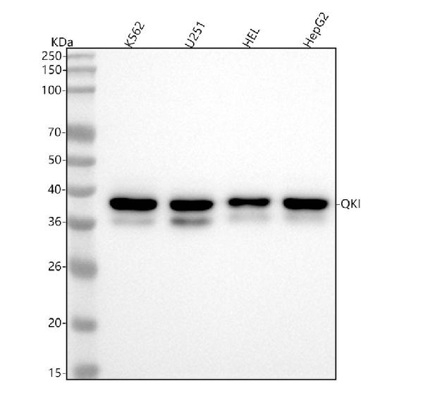
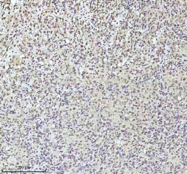
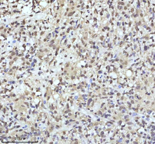
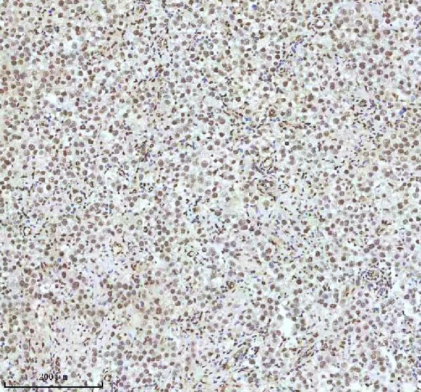
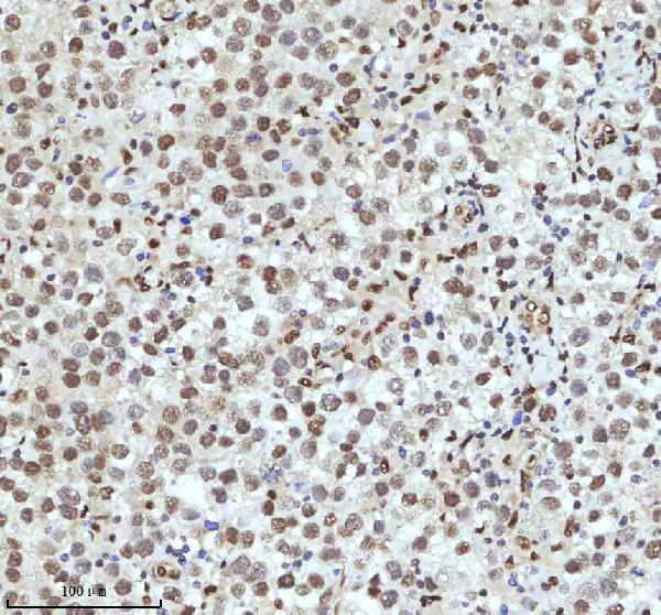
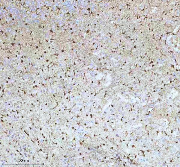
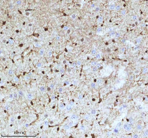
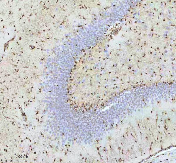
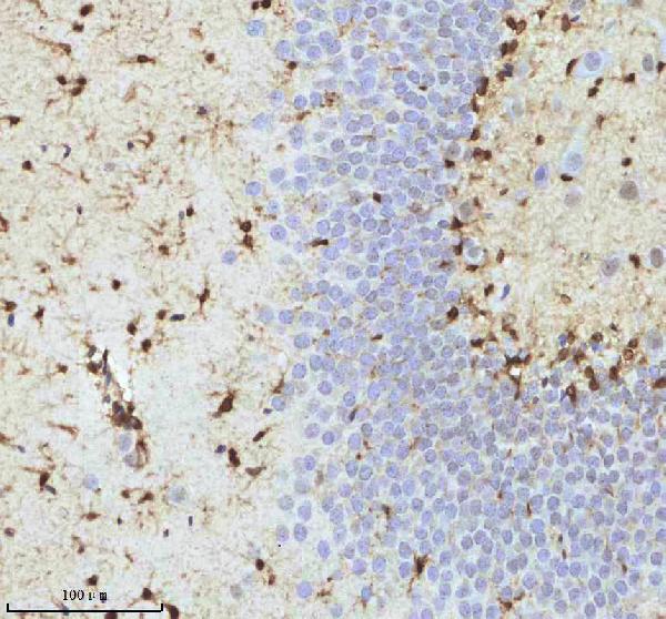
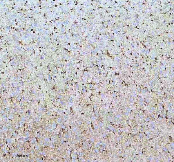
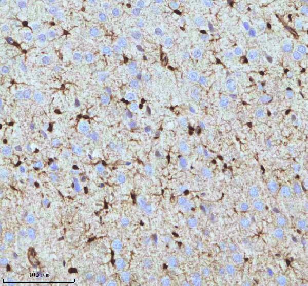
| Description | Anti-QK1 Rabbit Monoclonal Antibody . Tested in WB, IHC, ICC/IF, IP applications. This antibody reacts with Human, Mouse. |
| Gene ID | 9444 |
|---|---|
| Other Names | KH domain-containing RNA-binding protein QKI, Protein quaking, Hqk, HqkI, QKI {ECO:0000303|PubMed:16342280, ECO:0000312|HGNC:HGNC:21100} |
| Calculated MW | 38 kDa |
| Application Details | WB 1:500-1:2000 IHC 1:50-1:200 ICC/IF 1:50-1:200 IP 1:50 |
| Contents | Rabbit IgG in phosphate buffered saline, pH 7.4, 150mM NaCl, 0.02% sodium azide and 50% glycerol, 0.4-0.5mg/ml BSA. |
| Clone Names | Clone: 26Q56 |
| Immunogen | A synthesized peptide derived from QK1 |
| Purification | Affinity-chromatography |
| Storage | Store at -20°C for one year. For short term storage and frequent use, store at 4°C for up to one month. Avoid repeated freeze-thaw cycles. |
| Name | QKI {ECO:0000303|PubMed:16342280, ECO:0000312|HGNC:HGNC:21100} |
|---|---|
| Function | RNA reader protein, which recognizes and binds specific RNAs, thereby regulating RNA metabolic processes, such as pre-mRNA splicing, circular RNA (circRNA) formation, mRNA export, mRNA stability and/or translation (PubMed:22398723, PubMed:23630077, PubMed:25768908, PubMed:27029405, PubMed:31331967, PubMed:37379838). Involved in various cellular processes, such as mRNA storage into stress granules, apoptosis, lipid deposition, interferon response, glial cell fate and development (PubMed:25768908, PubMed:31829086, PubMed:34428287, PubMed:37379838). Binds to the 5'-NACUAAY-N(1,20)-UAAY-3' RNA core sequence (PubMed:23630077). Acts as a mRNA modification reader that specifically recognizes and binds mRNA transcripts modified by internal N(7)-methylguanine (m7G) (PubMed:37379838). Promotes the formation of circular RNAs (circRNAs) during the epithelial to mesenchymal transition and in cardiomyocytes: acts by binding to sites flanking circRNA-forming exons (PubMed:25768908). CircRNAs are produced by back- splicing circularization of pre-mRNAs (PubMed:25768908). Plays a central role in myelinization via 3 distinct mechanisms (PubMed:16641098). First, acts by protecting and promoting stability of target mRNAs such as MBP, SIRT2 and CDKN1B, which promotes oligodendrocyte differentiation (By similarity). Second, participates in mRNA transport by regulating the nuclear export of MBP mRNA (By similarity). Finally, indirectly regulates mRNA splicing of MAG pre- mRNA during oligodendrocyte differentiation by acting as a negative regulator of MAG exon 12 alternative splicing: acts by binding to HNRNPA1 mRNA splicing factor, preventing its translation (By similarity). Involved in microglia differentiation and remyelination by regulating microexon alternative splicing of the Rho GTPase pathway (By similarity). Involved in macrophage differentiation: promotes monocyte differentiation by regulating pre-mRNA splicing in naive peripheral blood monocytes (PubMed:27029405). Acts as an important regulator of muscle development: required for the contractile function of cardiomyocytes by regulating alternative splicing of cardiomyocyte transcripts (By similarity). Acts as a negative regulator of thermogenesis by decreasing stability, nuclear export and translation of mRNAs encoding PPARGC1A and UCP1 (By similarity). Also required for visceral endoderm function and blood vessel development (By similarity). May also play a role in smooth muscle development (PubMed:31331967). In addition to its RNA-binding activity, also acts as a nuclear transcription coactivator for SREBF2/SREBP2 (By similarity). |
| Cellular Location | Nucleus. Cytoplasm [Isoform QKI6]: Cytoplasm, cytosol. Nucleus Note=Localizes predominantly in the cytoplasm and at lower levels in nucleus. |
| Tissue Location | Expressed in the frontal cortex of brain. Down- regulated in the brain of schizophrenic patients |
Research Areas
Citations (0)

Thousands of laboratories across the world have published research that depended on the performance of antibodies from Abcepta to advance their research. Check out links to articles that cite our products in major peer-reviewed journals, organized by research category.
Submit your citation using an Abcepta antibody to
info@abcepta.com, and receive a free "I Love Antibodies" mug.
info@abcepta.com, and receive a free "I Love Antibodies" mug.

Application Protocols
Provided below are standard protocols that you may find useful for product applications.
Abcepta welcomes feedback from its customers.
If you have used an Abcepta product and would like to share how it has performed, please click on the "Submit Review" button and provide the requested information. Our staff will examine and post your review and contact you if needed.
If you have any additional inquiries please email technical services at tech@abcepta.com.
$ 370.00
Cat# ABO16077
Ordering Information
United States
AlbaniaAustraliaAustriaBelgiumBosnia & HerzegovinaBrazilBulgariaCanadaCentral AmericaChinaCroatiaCyprusCzech RepublicDenmarkEstoniaFinlandFranceGermanyGreeceHong KongHungaryIcelandIndiaIndonesiaIrelandIsraelItalyJapanLatviaLithuaniaLuxembourgMacedoniaMalaysiaMaltaMexicoNetherlandsNew ZealandNorwayPakistanPolandPortugalRomaniaSerbiaSingaporeSlovakiaSloveniaSouth AfricaSouth KoreaSpainSwedenSwitzerlandTaiwanTurkeyUnited KingdomUnited StatesVietnamWorldwideOthers
USA Headquarters
(888) 735-7227 / (858) 622-0099 or (858) 875-1900
Other Products
Shipping Information
Domestic orders (in stock items)
Shipped out the same day. Orders placed after 1 PM (PST) will ship out the next business day.
International orders
Contact your local distributors
