


 Foundational characteristics of cancer include proliferation, angiogenesis, migration, evasion of apoptosis, and cellular immortality. Find key markers for these cellular processes and antibodies to detect them.
 The SUMOplot™ Analysis Program predicts and scores sumoylation sites in your protein. SUMOylation is a post-translational modification involved in various cellular processes, such as nuclear-cytosolic transport, transcriptional regulation, apoptosis, protein stability, response to stress, and progression through the cell cycle.
 The Autophagy Receptor Motif Plotter predicts and scores autophagy receptor binding sites in your protein. Identifying proteins connected to this pathway is critical to understanding the role of autophagy in physiological as well as pathological processes such as development, differentiation, neurodegenerative diseases, stress, infection, and cancer.
> home > Products > Primary Antibodies > Signal Transduction > Anti-IGF2BP2 Rabbit Monoclonal Antibody
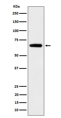
Anti-IGF2BP2 Rabbit Monoclonal Antibody
- SPECIFICATION
- CITATIONS
- PROTOCOLS
- BACKGROUND

Application
| WB, IHC |
|---|---|
| Primary Accession | Q9Y6M1 |
| Host | Rabbit |
| Isotype | IgG |
| Reactivity | Rat, Human, Mouse |
| Clonality | Monoclonal |
| Format | Liquid |
| Description | Anti-IGF2BP2 Rabbit Monoclonal Antibody . Tested in WB, IHC applications. This antibody reacts with Human, Mouse, Rat. |
| Gene ID | 10644 |
|---|---|
| Other Names | Insulin-like growth factor 2 mRNA-binding protein 2, IGF2 mRNA-binding protein 2, IMP-2, Hepatocellular carcinoma autoantigen p62, IGF-II mRNA-binding protein 2, VICKZ family member 2, IGF2BP2, IMP2, VICKZ2 |
| Calculated MW | 66 kDa |
| Application Details | WB 1:500-1:2000 IHC 1:50-1:200 |
| Contents | Rabbit IgG in phosphate buffered saline, pH 7.4, 150mM NaCl, 0.02% sodium azide and 50% glycerol, 0.4-0.5mg/ml BSA. |
| Clone Names | Clone: 26I86 |
| Immunogen | A synthesized peptide derived from human IGF2BP2 |
| Purification | Affinity-chromatography |
| Storage | Store at -20°C for one year. For short term storage and frequent use, store at 4°C for up to one month. Avoid repeated freeze-thaw cycles. |
| Name | IGF2BP2 |
|---|---|
| Synonyms | IMP2, VICKZ2 |
| Function | RNA-binding factor that recruits target transcripts to cytoplasmic protein-RNA complexes (mRNPs). This transcript 'caging' into mRNPs allows mRNA transport and transient storage. It also modulates the rate and location at which target transcripts encounter the translational apparatus and shields them from endonuclease attacks or microRNA-mediated degradation (By similarity). Preferentially binds to N6-methyladenosine (m6A)-containing mRNAs and increases their stability (PubMed:29476152). Binds to the 5'-UTR of the insulin-like growth factor 2 (IGF2) mRNAs (PubMed:9891060). Binding is isoform- specific. Binds to beta-actin/ACTB and MYC transcripts. Increases MYC mRNA stability by binding to the coding region instability determinant (CRD) and binding is enhanced by m6A-modification of the CRD (PubMed:29476152). |
| Cellular Location | Nucleus. Cytoplasm. Cytoplasm, P-body. Cytoplasm, Stress granule. Note=Localized in cytoplasmic mRNP granules containing untranslated mRNAs. Localizes at the connecting piece and the tail of the spermatozoa. In response to cellular stress, such as oxidative stress, recruited to stress granules |
| Tissue Location | Expressed in oocytes, granulosa cells of small and growing follicles, Leydig cells, spermatogonia and semen (at protein level). Expressed in testicular cancer (at protein level). Expressed weakly in heart, placenta, skeletal muscle, bone marrow, colon, kidney, salivary glands, testis and pancreas. Detected in fetal liver, fetal ovary, gonocytes and interstitial cells of the testis |
Research Areas
Citations (0)

Thousands of laboratories across the world have published research that depended on the performance of antibodies from Abcepta to advance their research. Check out links to articles that cite our products in major peer-reviewed journals, organized by research category.
Submit your citation using an Abcepta antibody to
info@abcepta.com, and receive a free "I Love Antibodies" mug.
info@abcepta.com, and receive a free "I Love Antibodies" mug.

Application Protocols
Provided below are standard protocols that you may find useful for product applications.
Abcepta welcomes feedback from its customers.
If you have used an Abcepta product and would like to share how it has performed, please click on the "Submit Review" button and provide the requested information. Our staff will examine and post your review and contact you if needed.
If you have any additional inquiries please email technical services at tech@abcepta.com.
$ 370.00
Cat# ABO16107
Ordering Information
United States
AlbaniaAustraliaAustriaBelgiumBosnia & HerzegovinaBrazilBulgariaCanadaCentral AmericaChinaCroatiaCyprusCzech RepublicDenmarkEstoniaFinlandFranceGermanyGreeceHong KongHungaryIcelandIndiaIndonesiaIrelandIsraelItalyJapanLatviaLithuaniaLuxembourgMacedoniaMalaysiaMaltaMexicoNetherlandsNew ZealandNorwayPakistanPolandPortugalRomaniaSerbiaSingaporeSlovakiaSloveniaSouth AfricaSouth KoreaSpainSwedenSwitzerlandTaiwanTurkeyUnited KingdomUnited StatesVietnamWorldwideOthers
USA Headquarters
(888) 735-7227 / (858) 622-0099 or (858) 875-1900
Other Products
Shipping Information
Domestic orders (in stock items)
Shipped out the same day. Orders placed after 1 PM (PST) will ship out the next business day.
International orders
Contact your local distributors
