


 Foundational characteristics of cancer include proliferation, angiogenesis, migration, evasion of apoptosis, and cellular immortality. Find key markers for these cellular processes and antibodies to detect them.
 The SUMOplot™ Analysis Program predicts and scores sumoylation sites in your protein. SUMOylation is a post-translational modification involved in various cellular processes, such as nuclear-cytosolic transport, transcriptional regulation, apoptosis, protein stability, response to stress, and progression through the cell cycle.
 The Autophagy Receptor Motif Plotter predicts and scores autophagy receptor binding sites in your protein. Identifying proteins connected to this pathway is critical to understanding the role of autophagy in physiological as well as pathological processes such as development, differentiation, neurodegenerative diseases, stress, infection, and cancer.
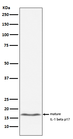
Anti-IL-1 beta Rabbit Monoclonal Antibody
- SPECIFICATION
- CITATIONS
- PROTOCOLS
- BACKGROUND

Application
| WB, IP, FC |
|---|---|
| Primary Accession | P10749 |
| Host | Rabbit |
| Isotype | IgG |
| Reactivity | Mouse |
| Clonality | Monoclonal |
| Format | Liquid |
| Description | Anti-IL-1 beta Rabbit Monoclonal Antibody . Tested in WB, IP, Flow Cytometry applications. This antibody reacts with Mouse. |
| Gene ID | 16176 |
|---|---|
| Other Names | Interleukin-1 beta, IL-1 beta, Il1b |
| Calculated MW | 31 kDa, 28 kDa, 17 kDa |
| Application Details | WB 1:500-1:2000 IP 1:50 FC 1:50 |
| Contents | Rabbit IgG in phosphate buffered saline, pH 7.4, 150mM NaCl, 0.02% sodium azide and 50% glycerol, 0.4-0.5mg/ml BSA. |
| Clone Names | Clone: 28I10 |
| Immunogen | A synthesized peptide derived from human IL-1 beta |
| Purification | Affinity-chromatography |
| Storage | Store at -20°C for one year. For short term storage and frequent use, store at 4°C for up to one month. Avoid repeated freeze-thaw cycles. |
| Name | Il1b |
|---|---|
| Function | Potent pro-inflammatory cytokine. Initially discovered as the major endogenous pyrogen, induces prostaglandin synthesis, neutrophil influx and activation, T-cell activation and cytokine production, B- cell activation and antibody production, and fibroblast proliferation and collagen production. Promotes Th17 differentiation of T-cells. Synergizes with IL12/interleukin-12 to induce IFNG synthesis from T- helper 1 (Th1) cells. Plays a role in angiogenesis by inducing VEGF production synergistically with TNF and IL6. Involved in transduction of inflammation downstream of pyroptosis: its mature form is specifically released in the extracellular milieu by passing through the gasdermin-D (GSDMD) pore. |
| Cellular Location | Cytoplasm, cytosol. Secreted Lysosome {ECO:0000250|UniProtKB:P01584}. Secreted, extracellular exosome. Note=The precursor is cytosolic In response to inflammasome-activating signals, such as ATP for NLRP3 inflammasome or bacterial flagellin for NLRC4 inflammasome, cleaved and secreted. Mature form is secreted and released in the extracellular milieu by passing through the gasdermin-D (GSDMD) pore. In contrast, the precursor form is not released, due to the presence of an acidic region that is proteolytically removed by CASP1 during maturation. The secretion is dependent on protein unfolding and facilitated by the cargo receptor TMED10. {ECO:0000250|UniProtKB:P01584} |
| Tissue Location | Expressed in activated macrophages (at protein level). |
Research Areas
Citations (0)

Thousands of laboratories across the world have published research that depended on the performance of antibodies from Abcepta to advance their research. Check out links to articles that cite our products in major peer-reviewed journals, organized by research category.
Submit your citation using an Abcepta antibody to
info@abcepta.com, and receive a free "I Love Antibodies" mug.
info@abcepta.com, and receive a free "I Love Antibodies" mug.

Application Protocols
Provided below are standard protocols that you may find useful for product applications.
Abcepta welcomes feedback from its customers.
If you have used an Abcepta product and would like to share how it has performed, please click on the "Submit Review" button and provide the requested information. Our staff will examine and post your review and contact you if needed.
If you have any additional inquiries please email technical services at tech@abcepta.com.
$ 370.00
Cat# ABO16231
Ordering Information
United States
AlbaniaAustraliaAustriaBelgiumBosnia & HerzegovinaBrazilBulgariaCanadaCentral AmericaChinaCroatiaCyprusCzech RepublicDenmarkEstoniaFinlandFranceGermanyGreeceHong KongHungaryIcelandIndiaIndonesiaIrelandIsraelItalyJapanLatviaLithuaniaLuxembourgMacedoniaMalaysiaMaltaMexicoNetherlandsNew ZealandNorwayPakistanPolandPortugalRomaniaSerbiaSingaporeSlovakiaSloveniaSouth AfricaSouth KoreaSpainSwedenSwitzerlandTaiwanTurkeyUnited KingdomUnited StatesVietnamWorldwideOthers
USA Headquarters
(888) 735-7227 / (858) 622-0099 or (858) 875-1900
Other Products
Shipping Information
Domestic orders (in stock items)
Shipped out the same day. Orders placed after 1 PM (PST) will ship out the next business day.
International orders
Contact your local distributors
