


 Foundational characteristics of cancer include proliferation, angiogenesis, migration, evasion of apoptosis, and cellular immortality. Find key markers for these cellular processes and antibodies to detect them.
 The SUMOplot™ Analysis Program predicts and scores sumoylation sites in your protein. SUMOylation is a post-translational modification involved in various cellular processes, such as nuclear-cytosolic transport, transcriptional regulation, apoptosis, protein stability, response to stress, and progression through the cell cycle.
 The Autophagy Receptor Motif Plotter predicts and scores autophagy receptor binding sites in your protein. Identifying proteins connected to this pathway is critical to understanding the role of autophagy in physiological as well as pathological processes such as development, differentiation, neurodegenerative diseases, stress, infection, and cancer.
Anti-AK2 Rabbit Monoclonal Antibody
- SPECIFICATION
- CITATIONS
- PROTOCOLS
- BACKGROUND

Application
| WB, IHC, IF, ICC, IP |
|---|---|
| Primary Accession | P54819 |
| Host | Rabbit |
| Isotype | IgG |
| Reactivity | Rat, Human, Mouse |
| Clonality | Monoclonal |
| Format | Liquid |
| Description | Anti-AK2 Rabbit Monoclonal Antibody . Tested in WB, IHC, ICC/IF, IP applications. This antibody reacts with Human, Mouse, Rat. |
| Gene ID | 204 |
|---|---|
| Other Names | Adenylate kinase 2, mitochondrial {ECO:0000255|HAMAP-Rule:MF_03168}, AK 2 {ECO:0000255|HAMAP-Rule:MF_03168}, 2.7.4.3 {ECO:0000255|HAMAP-Rule:MF_03168}, ATP-AMP transphosphorylase 2 {ECO:0000255|HAMAP-Rule:MF_03168}, ATP:AMP phosphotransferase {ECO:0000255|HAMAP-Rule:MF_03168}, Adenylate monophosphate kinase {ECO:0000255|HAMAP-Rule:MF_03168}, Adenylate kinase 2, mitochondrial, N-terminally processed {ECO:0000255|HAMAP-Rule:MF_03168}, AK2 {ECO:0000255|HAMAP-Rule:MF_03168}, ADK2 |
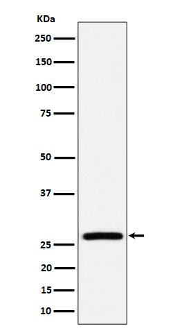
| Calculated MW | 26 kDa |
| Application Details | WB 1:500-1:2000 IHC 1:50-1:200 ICC/IF 1:50-1:200 IP 1:50 |
| Contents | Rabbit IgG in phosphate buffered saline, pH 7.4, 150mM NaCl, 0.02% sodium azide and 50% glycerol, 0.4-0.5mg/ml BSA. |
| Clone Names | Clone: 29A42 |
| Immunogen | A synthesized peptide derived from human AK2 |
| Purification | Affinity-chromatography |
| Storage | Store at -20°C for one year. For short term storage and frequent use, store at 4°C for up to one month. Avoid repeated freeze-thaw cycles. |
| Name | AK2 {ECO:0000255|HAMAP-Rule:MF_03168} |
|---|---|
| Synonyms | ADK2 |
| Function | Catalyzes the reversible transfer of the terminal phosphate group between ATP and AMP. Plays an important role in cellular energy homeostasis and in adenine nucleotide metabolism. Adenylate kinase activity is critical for regulation of the phosphate utilization and the AMP de novo biosynthesis pathways. Plays a key role in hematopoiesis. |
| Cellular Location | Mitochondrion intermembrane space {ECO:0000255|HAMAP-Rule:MF_03168} |
| Tissue Location | Present in most tissues. Present at high level in heart, liver and kidney, and at low level in brain, skeletal muscle and skin. Present in thrombocytes but not in erythrocytes, which lack mitochondria. Present in all nucleated cell populations from blood, while AK1 is mostly absent. In spleen and lymph nodes, mononuclear cells lack AK1, whereas AK2 is readily detectable. These results indicate that leukocytes may be susceptible to defects caused by the lack of AK2, as they do not express AK1 in sufficient amounts to compensate for the AK2 functional deficits (at protein level) |
Research Areas
Citations (0)

Thousands of laboratories across the world have published research that depended on the performance of antibodies from Abcepta to advance their research. Check out links to articles that cite our products in major peer-reviewed journals, organized by research category.
Submit your citation using an Abcepta antibody to
info@abcepta.com, and receive a free "I Love Antibodies" mug.
info@abcepta.com, and receive a free "I Love Antibodies" mug.

Application Protocols
Provided below are standard protocols that you may find useful for product applications.
Abcepta welcomes feedback from its customers.
If you have used an Abcepta product and would like to share how it has performed, please click on the "Submit Review" button and provide the requested information. Our staff will examine and post your review and contact you if needed.
If you have any additional inquiries please email technical services at tech@abcepta.com.
$ 370.00
Cat# ABO16406
Ordering Information
United States
AlbaniaAustraliaAustriaBelgiumBosnia & HerzegovinaBrazilBulgariaCanadaCentral AmericaChinaCroatiaCyprusCzech RepublicDenmarkEstoniaFinlandFranceGermanyGreeceHong KongHungaryIcelandIndiaIndonesiaIrelandIsraelItalyJapanLatviaLithuaniaLuxembourgMacedoniaMalaysiaMaltaMexicoNetherlandsNew ZealandNorwayPakistanPolandPortugalRomaniaSerbiaSingaporeSlovakiaSloveniaSouth AfricaSouth KoreaSpainSwedenSwitzerlandTaiwanTurkeyUnited KingdomUnited StatesVietnamWorldwideOthers
USA Headquarters
(888) 735-7227 / (858) 622-0099 or (858) 875-1900
Other Products
Shipping Information
Domestic orders (in stock items)
Shipped out the same day. Orders placed after 1 PM (PST) will ship out the next business day.
International orders
Contact your local distributors
