


 Foundational characteristics of cancer include proliferation, angiogenesis, migration, evasion of apoptosis, and cellular immortality. Find key markers for these cellular processes and antibodies to detect them.
 The SUMOplot™ Analysis Program predicts and scores sumoylation sites in your protein. SUMOylation is a post-translational modification involved in various cellular processes, such as nuclear-cytosolic transport, transcriptional regulation, apoptosis, protein stability, response to stress, and progression through the cell cycle.
 The Autophagy Receptor Motif Plotter predicts and scores autophagy receptor binding sites in your protein. Identifying proteins connected to this pathway is critical to understanding the role of autophagy in physiological as well as pathological processes such as development, differentiation, neurodegenerative diseases, stress, infection, and cancer.
> home > Products > Primary Antibodies > Antibody Collections > Pancreas negative > Anti-TMEM43 Rabbit Monoclonal Antibody
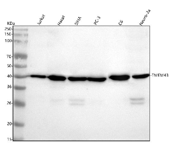
Anti-TMEM43 Rabbit Monoclonal Antibody
- SPECIFICATION
- CITATIONS
- PROTOCOLS
- BACKGROUND

Application
| WB, IHC |
|---|---|
| Primary Accession | Q9BTV4 |
| Host | Rabbit |
| Isotype | IgG |
| Reactivity | Rat, Human, Mouse |
| Clonality | Monoclonal |
| Format | Liquid |
| Description | Anti-TMEM43 Rabbit Monoclonal Antibody . Tested in WB, IHC applications. This antibody reacts with Human, Mouse, Rat. |
| Gene ID | 79188 |
|---|---|
| Other Names | Transmembrane protein 43, Protein LUMA, TMEM43 |
| Calculated MW | 45 kDa |
| Application Details | WB 1:500-1:2000 IHC 1:50-1:200 |
| Contents | Rabbit IgG in phosphate buffered saline, pH 7.4, 150mM NaCl, 0.02% sodium azide and 50% glycerol, 0.4-0.5mg/ml BSA. |
| Clone Names | Clone: 30T30 |
| Immunogen | A synthesized peptide derived from human TMEM43 |
| Purification | Affinity-chromatography |
| Storage | Store at -20°C for one year. For short term storage and frequent use, store at 4°C for up to one month. Avoid repeated freeze-thaw cycles. |
| Name | TMEM43 |
|---|---|
| Function | May have an important role in maintaining nuclear envelope structure by organizing protein complexes at the inner nuclear membrane. Required for retaining emerin at the inner nuclear membrane (By similarity). Plays a role in the modulation of innate immune signaling through the cGAS-STING pathway by interacting with RNF26 (PubMed:32614325). In addition, functions as a critical signaling component in mediating NF-kappa-B activation by acting downstream of EGFR and upstream of CARD10 (PubMed:27991920). Contributes to passive conductance current in cochlear glia-like supporting cells, mediated by gap junctions and necessary for hearing and speech discrimination (PubMed:34050020). |
| Cellular Location | Endoplasmic reticulum membrane. Nucleus inner membrane; Multi-pass membrane protein. Cell membrane Note=Retained in the inner nuclear membrane through interaction with EMD and A- and B-lamins. The N- and C-termini are oriented towards the nucleoplasm. The majority of the hydrophilic domain resides in the endoplasmic reticulum lumen (By similarity). |
| Tissue Location | Highest expression in placenta. Also found at lower levels in heart, ovary, spleen, small intestine, thymus, prostate and testis. |
Research Areas
Citations (0)

Thousands of laboratories across the world have published research that depended on the performance of antibodies from Abcepta to advance their research. Check out links to articles that cite our products in major peer-reviewed journals, organized by research category.
Submit your citation using an Abcepta antibody to
info@abcepta.com, and receive a free "I Love Antibodies" mug.
info@abcepta.com, and receive a free "I Love Antibodies" mug.

Application Protocols
Provided below are standard protocols that you may find useful for product applications.
Abcepta welcomes feedback from its customers.
If you have used an Abcepta product and would like to share how it has performed, please click on the "Submit Review" button and provide the requested information. Our staff will examine and post your review and contact you if needed.
If you have any additional inquiries please email technical services at tech@abcepta.com.
$ 370.00
Cat# ABO16494
Ordering Information
United States
AlbaniaAustraliaAustriaBelgiumBosnia & HerzegovinaBrazilBulgariaCanadaCentral AmericaChinaCroatiaCyprusCzech RepublicDenmarkEstoniaFinlandFranceGermanyGreeceHong KongHungaryIcelandIndiaIndonesiaIrelandIsraelItalyJapanLatviaLithuaniaLuxembourgMacedoniaMalaysiaMaltaMexicoNetherlandsNew ZealandNorwayPakistanPolandPortugalRomaniaSerbiaSingaporeSlovakiaSloveniaSouth AfricaSouth KoreaSpainSwedenSwitzerlandTaiwanTurkeyUnited KingdomUnited StatesVietnamWorldwideOthers
USA Headquarters
(888) 735-7227 / (858) 622-0099 or (858) 875-1900
Other Products
Shipping Information
Domestic orders (in stock items)
Shipped out the same day. Orders placed after 1 PM (PST) will ship out the next business day.
International orders
Contact your local distributors
