


 Foundational characteristics of cancer include proliferation, angiogenesis, migration, evasion of apoptosis, and cellular immortality. Find key markers for these cellular processes and antibodies to detect them.
 The SUMOplot™ Analysis Program predicts and scores sumoylation sites in your protein. SUMOylation is a post-translational modification involved in various cellular processes, such as nuclear-cytosolic transport, transcriptional regulation, apoptosis, protein stability, response to stress, and progression through the cell cycle.
 The Autophagy Receptor Motif Plotter predicts and scores autophagy receptor binding sites in your protein. Identifying proteins connected to this pathway is critical to understanding the role of autophagy in physiological as well as pathological processes such as development, differentiation, neurodegenerative diseases, stress, infection, and cancer.
> home > Products > Primary Antibodies > Signal Transduction > Anti-Clusterin Rabbit Monoclonal Antibody
Anti-Clusterin Rabbit Monoclonal Antibody
- SPECIFICATION
- CITATIONS
- PROTOCOLS
- BACKGROUND

Application
| WB, IHC, IP |
|---|---|
| Primary Accession | Q06890 |
| Host | Rabbit |
| Isotype | IgG |
| Reactivity | Rat, Mouse |
| Clonality | Monoclonal |
| Format | Liquid |
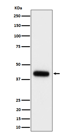
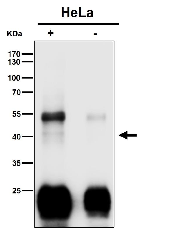
| Description | Anti-Clusterin Rabbit Monoclonal Antibody . Tested in WB, IHC, IP applications. This antibody reacts with Mouse, Rat. |
| Gene ID | 12759 |
|---|---|
| Other Names | Clusterin, Apolipoprotein J, Apo-J, Clustrin {ECO:0000303|Ref.4}, Sulfated glycoprotein 2, SGP-2, Clusterin beta chain, Clusterin alpha chain, Clu {ECO:0000312|MGI:MGI:88423} |
| Calculated MW | 32-42 kDa, 65 kDa, 75 kDa |
| Application Details | WB 1:500-1:2000 IHC 1:50-1:200 IP 1:50 |
| Contents | Rabbit IgG in phosphate buffered saline, pH 7.4, 150mM NaCl, 0.02% sodium azide and 50% glycerol, 0.4-0.5mg/ml BSA. |
| Clone Names | Clone: 30C68 |
| Immunogen | A synthesized peptide derived from human Clusterin |
| Purification | Affinity-chromatography |
| Storage | Store at -20°C for one year. For short term storage and frequent use, store at 4°C for up to one month. Avoid repeated freeze-thaw cycles. |
| Name | Clu {ECO:0000312|MGI:MGI:88423} |
|---|---|
| Function | Functions as extracellular chaperone that prevents aggregation of non native proteins. Prevents stress-induced aggregation of blood plasma proteins (By similarity). Inhibits formation of amyloid fibrils by APP, APOC2, B2M, CALCA, CSN3, SNCA and aggregation-prone LYZ variants (in vitro) (PubMed:14741101). Does not require ATP. Maintains partially unfolded proteins in a state appropriate for subsequent refolding by other chaperones, such as HSPA8/HSC70. Does not refold proteins by itself. Binding to cell surface receptors triggers internalization of the chaperone-client complex and subsequent lysosomal or proteasomal degradation. When secreted, protects cells against apoptosis and against cytolysis by complement: inhibits assembly of the complement membrane attack complex (MAC) by preventing polymerization of C9 pore component of the MAC complex. Intracellular forms interact with ubiquitin and SCF (SKP1-CUL1-F-box protein) E3 ubiquitin-protein ligase complexes and promote the ubiquitination and subsequent proteasomal degradation of target proteins. Promotes proteasomal degradation of COMMD1 and IKBKB. Modulates NF-kappa-B transcriptional activity (By similarity). Following stress, promotes apoptosis (PubMed:12551933). Inhibits apoptosis when associated with the mitochondrial membrane by interference with BAX-dependent release of cytochrome c into the cytoplasm. Plays a role in the regulation of cell proliferation. Following ER stress, suppresses stress-induced apoptosis by stabilizing mitochondrial membrane integrity through interaction with HSPA5. When secreted, does not affect caspase or BAX- mediated intrinsic apoptosis and TNF-induced NF-kappa-B-activity (By similarity). When secreted, acts as an important modulator during neuronal differentiation through interaction with STMN3 (By similarity). Plays a role in the clearance of immune complexes that arise during cell injury (PubMed:11865066). |
| Cellular Location | Secreted. Nucleus. Cytoplasm Mitochondrion membrane {ECO:0000250|UniProtKB:P10909}; Peripheral membrane protein {ECO:0000250|UniProtKB:P10909}; Cytoplasmic side {ECO:0000250|UniProtKB:P10909}. Cytoplasm, cytosol. Microsome {ECO:0000250|UniProtKB:P10909}. Endoplasmic reticulum {ECO:0000250|UniProtKB:P10909}. Mitochondrion {ECO:0000250|UniProtKB:P10909}. Mitochondrion membrane {ECO:0000250|UniProtKB:P10909}. Cytoplasm, perinuclear region {ECO:0000250|UniProtKB:P05371}. Cytoplasmic vesicle, secretory vesicle, chromaffin granule {ECO:0000250|UniProtKB:P10909}. Note=Can retrotranslocate from the secretory compartments to the cytosol upon cellular stress. Detected in perinuclear foci that may be aggresomes containing misfolded, ubiquitinated proteins. Detected at the mitochondrion membrane upon induction of apoptosis. Under ER stress, a immaturely glycosylated pre-secreted form retrotranslocates from the endoplasmic reticulum (ER)-Golgi network to the cytoplasm to localize in the mitochondria through HSPA5 interaction. ER stress reduces secretion. Under the stress, minor amounts of non-secreted forms accumulate in cytoplasm. {ECO:0000250|UniProtKB:P10909} |
| Tissue Location | Most abundant in stomach, liver, brain, and testis, with intermediate levels in heart, ovary and kidney |
Research Areas
Citations (0)

Thousands of laboratories across the world have published research that depended on the performance of antibodies from Abcepta to advance their research. Check out links to articles that cite our products in major peer-reviewed journals, organized by research category.
Submit your citation using an Abcepta antibody to
info@abcepta.com, and receive a free "I Love Antibodies" mug.
info@abcepta.com, and receive a free "I Love Antibodies" mug.

Application Protocols
Provided below are standard protocols that you may find useful for product applications.
Abcepta welcomes feedback from its customers.
If you have used an Abcepta product and would like to share how it has performed, please click on the "Submit Review" button and provide the requested information. Our staff will examine and post your review and contact you if needed.
If you have any additional inquiries please email technical services at tech@abcepta.com.
$ 370.00
Cat# ABO16532
Ordering Information
United States
AlbaniaAustraliaAustriaBelgiumBosnia & HerzegovinaBrazilBulgariaCanadaCentral AmericaChinaCroatiaCyprusCzech RepublicDenmarkEstoniaFinlandFranceGermanyGreeceHong KongHungaryIcelandIndiaIndonesiaIrelandIsraelItalyJapanLatviaLithuaniaLuxembourgMacedoniaMalaysiaMaltaMexicoNetherlandsNew ZealandNorwayPakistanPolandPortugalRomaniaSerbiaSingaporeSlovakiaSloveniaSouth AfricaSouth KoreaSpainSwedenSwitzerlandTaiwanTurkeyUnited KingdomUnited StatesVietnamWorldwideOthers
USA Headquarters
(888) 735-7227 / (858) 622-0099 or (858) 875-1900
Other Products
Shipping Information
Domestic orders (in stock items)
Shipped out the same day. Orders placed after 1 PM (PST) will ship out the next business day.
International orders
Contact your local distributors
