


 Foundational characteristics of cancer include proliferation, angiogenesis, migration, evasion of apoptosis, and cellular immortality. Find key markers for these cellular processes and antibodies to detect them.
 The SUMOplot™ Analysis Program predicts and scores sumoylation sites in your protein. SUMOylation is a post-translational modification involved in various cellular processes, such as nuclear-cytosolic transport, transcriptional regulation, apoptosis, protein stability, response to stress, and progression through the cell cycle.
 The Autophagy Receptor Motif Plotter predicts and scores autophagy receptor binding sites in your protein. Identifying proteins connected to this pathway is critical to understanding the role of autophagy in physiological as well as pathological processes such as development, differentiation, neurodegenerative diseases, stress, infection, and cancer.
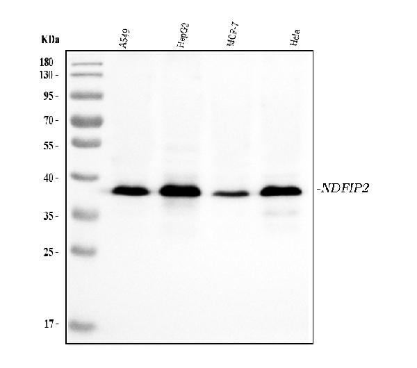
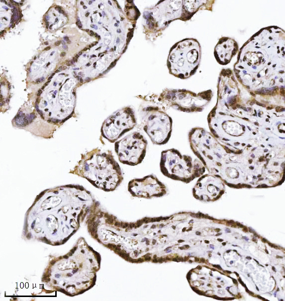
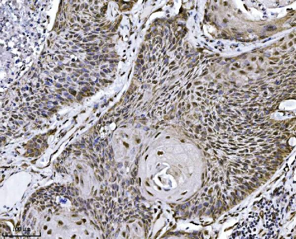
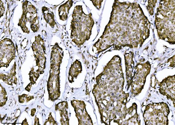
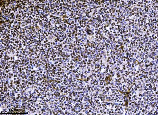
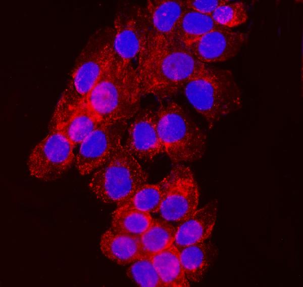
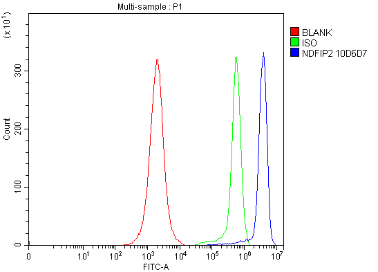
Anti-NDFIP2 Antibody Picoband™ (monoclonal, 10D6D7)
- SPECIFICATION
- CITATIONS
- PROTOCOLS
- BACKGROUND

Application
| WB, IHC, IF, ICC, FC |
|---|---|
| Primary Accession | Q9NV92 |
| Host | Mouse |
| Isotype | Mouse IgG2b |
| Reactivity | Human |
| Clonality | Monoclonal |
| Format | Lyophilized |
| Description | Anti-NDFIP2 Antibody Picoband™ (monoclonal, 10D6D7) . Tested in Flow Cytometry, IF, IHC, ICC, WB applications. This antibody reacts with Human. |
| Reconstitution | Adding 0.2 ml of distilled water will yield a concentration of 500 µg/ml. |
| Gene ID | 54602 |
|---|---|
| Other Names | NEDD4 family-interacting protein 2, NEDD4 WW domain-binding protein 5A, Putative MAPK-activating protein PM04/PM05/PM06/PM07, Putative NF-kappa-B-activating protein 413, NDFIP2, KIAA1165, N4WBP5A |
| Calculated MW | 39 kDa |
| Application Details | Western blot, 0.25-0.5 µg/ml, Human Immunohistochemistry(Paraffin-embedded Section), 2-5 µg/ml, Human Immunocytochemistry/Immunofluorescence, 5 µg/ml, Human Flow Cytometry, 1-3 µg/1x10^6 cells, Human |
| Contents | Each vial contains 4 mg Trehalose, 0.9 mg NaCl and 0.2 mg Na2HPO4. |
| Clone Names | Clone: 10D6D7 |
| Immunogen | E.coli-derived human NDFIP2 recombinant protein (Position: M16-L336). |
| Purification | Immunogen affinity purified. |
| Storage | At -20°C for one year from date of receipt. After reconstitution, at 4°C for one month. It can also be aliquotted and stored frozen at -20°C for six months. Avoid repeated freezing and thawing. |
| Name | NDFIP2 |
|---|---|
| Synonyms | KIAA1165, N4WBP5A |
| Function | Activates HECT domain-containing E3 ubiquitin-protein ligases, including ITCH, NEDD4, NEDD4L, SMURF2, WWP1 and WWP2, and consequently modulates the stability of their targets. As a result, may control many cellular processes. Recruits ITCH, NEDD4 and SMURF2 to endosomal membranes. Negatively regulates KCNH2 potassium channel activity by decreasing its cell-surface expression and interfering with channel maturation through recruitment of NEDD4L to the Golgi apparatus and multivesicular body where it mediates KCNH2 degradation (PubMed:26363003). May modulate EGFR signaling. Together with NDFIP1, limits the cytokine signaling and expansion of effector Th2 T-cells by promoting degradation of JAK1, probably by ITCH- and NEDD4L-mediated ubiquitination (By similarity). |
| Cellular Location | Endosome membrane; Multi-pass membrane protein. Golgi apparatus membrane. Endosome, multivesicular body membrane |
| Tissue Location | Expressed in brain, lung, heart, skeletal muscle, kidney, liver and placenta. |

Thousands of laboratories across the world have published research that depended on the performance of antibodies from Abcepta to advance their research. Check out links to articles that cite our products in major peer-reviewed journals, organized by research category.
info@abcepta.com, and receive a free "I Love Antibodies" mug.

Provided below are standard protocols that you may find useful for product applications.
Background
NEDD4 family-interacting protein 2 is a protein that in humans is encoded by the NDFIP2 gene. The NEDD4 family-interacting protein 1 (NDFIP1) belongs to a small group of evolutionarily conserved proteins with three transmembrane domains and is an integral Golgi membrane protein. It is a potential target for ubiquitination by the Nedd4 family of proteins. NDFIP1 is strongly expressed in surviving neurons following acute cortical brain injury, and overexpression in cultured cortical neurons increased survival following growth factor starvation, suggesting that NDFIP1 may play a role in neuronal survival. NDFIP1 and the related protein NDFIP2 are thought to interact with and regulate multiple components of the EGF and PTEN/Akt signaling pathways. Recent studies suggest that NDFIP1 may also play a role in Th17 differentiation by limiting the production of proinflammatory cytokines.
If you have used an Abcepta product and would like to share how it has performed, please click on the "Submit Review" button and provide the requested information. Our staff will examine and post your review and contact you if needed.
If you have any additional inquiries please email technical services at tech@abcepta.com.
Ordering Information
Other Products
Shipping Information
