


 Foundational characteristics of cancer include proliferation, angiogenesis, migration, evasion of apoptosis, and cellular immortality. Find key markers for these cellular processes and antibodies to detect them.
 The SUMOplot™ Analysis Program predicts and scores sumoylation sites in your protein. SUMOylation is a post-translational modification involved in various cellular processes, such as nuclear-cytosolic transport, transcriptional regulation, apoptosis, protein stability, response to stress, and progression through the cell cycle.
 The Autophagy Receptor Motif Plotter predicts and scores autophagy receptor binding sites in your protein. Identifying proteins connected to this pathway is critical to understanding the role of autophagy in physiological as well as pathological processes such as development, differentiation, neurodegenerative diseases, stress, infection, and cancer.
Anti-Erlin-2/ERLIN2 Antibody Picoband™ (monoclonal, 3H9A2)
- SPECIFICATION
- CITATIONS
- PROTOCOLS
- BACKGROUND

Application
| WB, IHC, IF, ICC, FC |
|---|---|
| Primary Accession | O94905 |
| Host | Mouse |
| Isotype | Mouse IgG2b |
| Reactivity | Human |
| Clonality | Monoclonal |
| Format | Lyophilized |
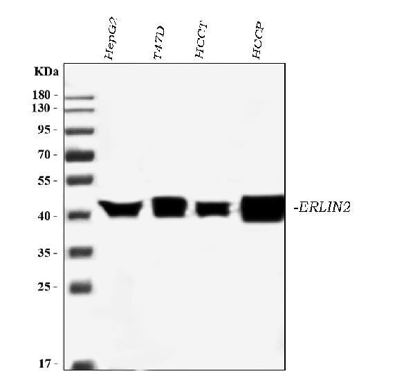
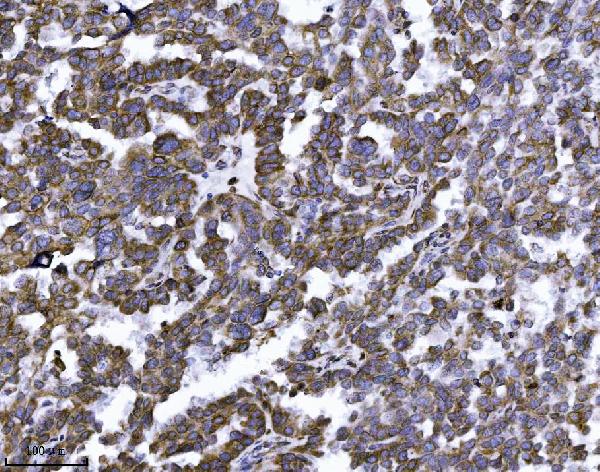
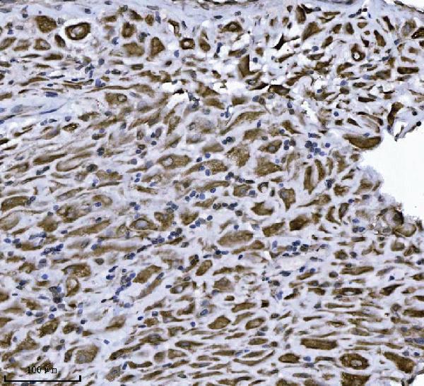
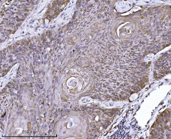
| Description | Anti-Erlin-2/ERLIN2 Antibody Picoband™ (monoclonal, 3H9A2) . Tested in Flow Cytometry, IF, IHC, ICC, WB applications. This antibody reacts with Human. |
| Reconstitution | Adding 0.2 ml of distilled water will yield a concentration of 500 µg/ml. |
| Gene ID | 11160 |
|---|---|
| Other Names | Erlin-2, Endoplasmic reticulum lipid raft-associated protein 2, Stomatin-prohibitin-flotillin-HflC/K domain-containing protein 2, SPFH domain-containing protein 2, ERLIN2, C8orf2, SPFH2 |
| Calculated MW | 43 kDa |
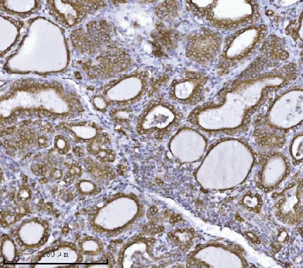
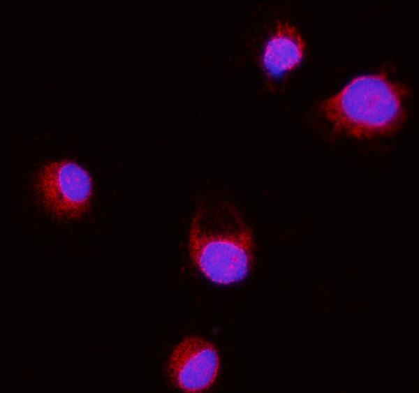
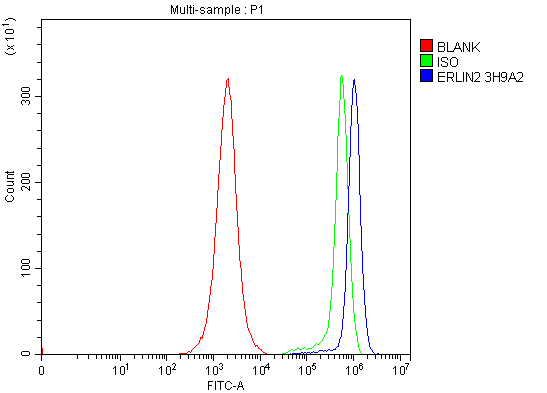
| Application Details | Western blot, 0.25-0.5 µg/ml, Human Immunohistochemistry(Paraffin-embedded Section), 2-5 µg/ml, Human Immunocytochemistry/Immunofluorescence, 5 µg/ml, Human Flow Cytometry, 1-3 µg/1x10^6 cells, Human |
| Contents | Each vial contains 4 mg Trehalose, 0.9 mg NaCl and 0.2 mg Na2HPO4. |
| Clone Names | Clone: 3H9A2 |
| Immunogen | E.coli-derived human Erlin-2/ERLIN2 recombinant protein (Position: D87-N339). |
| Purification | Immunogen affinity purified. |
| Storage | At -20°C for one year from date of receipt. After reconstitution, at 4°C for one month. It can also be aliquotted and stored frozen at -20°C for six months. Avoid repeated freezing and thawing. |
| Name | ERLIN2 |
|---|---|
| Synonyms | C8orf2, SPFH2 |
| Function | Component of the ERLIN1/ERLIN2 complex which mediates the endoplasmic reticulum-associated degradation (ERAD) of inositol 1,4,5- trisphosphate receptors (IP3Rs) such as ITPR1 (PubMed:17502376, PubMed:19240031). Promotes sterol-accelerated ERAD of HMGCR probably implicating an AMFR/gp78-containing ubiquitin ligase complex (PubMed:21343306). Involved in regulation of cellular cholesterol homeostasis by regulation the SREBP signaling pathway. May promote ER retention of the SCAP-SREBF complex (PubMed:24217618). |
| Cellular Location | Endoplasmic reticulum membrane; Single-pass type II membrane protein. Note=Associated with lipid raft-like domains of the endoplasmic reticulum membrane |
| Tissue Location | Ubiquitous.. |

Thousands of laboratories across the world have published research that depended on the performance of antibodies from Abcepta to advance their research. Check out links to articles that cite our products in major peer-reviewed journals, organized by research category.
info@abcepta.com, and receive a free "I Love Antibodies" mug.

Provided below are standard protocols that you may find useful for product applications.
Background
Erlin-2 is a protein that in humans is encoded by the ERLIN2 gene. This gene encodes a member of the SPFH domain-containing family of lipid raft-associated proteins. The encoded protein is localized to lipid rafts of the endoplasmic reticulum and plays a critical role in inositol 1,4,5-trisphosphate (IP3) signaling by mediating ER-associated degradation of activated IP3 receptors. Mutations in this gene are a cause of spastic paraplegia-18 (SPG18). Alternatively spliced transcript variants encoding multiple isoforms have been observed for this gene.
If you have used an Abcepta product and would like to share how it has performed, please click on the "Submit Review" button and provide the requested information. Our staff will examine and post your review and contact you if needed.
If you have any additional inquiries please email technical services at tech@abcepta.com.
Ordering Information
Other Products
Shipping Information
