


 Foundational characteristics of cancer include proliferation, angiogenesis, migration, evasion of apoptosis, and cellular immortality. Find key markers for these cellular processes and antibodies to detect them.
 The SUMOplot™ Analysis Program predicts and scores sumoylation sites in your protein. SUMOylation is a post-translational modification involved in various cellular processes, such as nuclear-cytosolic transport, transcriptional regulation, apoptosis, protein stability, response to stress, and progression through the cell cycle.
 The Autophagy Receptor Motif Plotter predicts and scores autophagy receptor binding sites in your protein. Identifying proteins connected to this pathway is critical to understanding the role of autophagy in physiological as well as pathological processes such as development, differentiation, neurodegenerative diseases, stress, infection, and cancer.
Anti-WDR1 Antibody Picoband™ (monoclonal, 5C11C8)
- SPECIFICATION
- CITATIONS
- PROTOCOLS
- BACKGROUND

Application
| WB, IHC |
|---|---|
| Primary Accession | O75083 |
| Host | Mouse |
| Isotype | Mouse IgG2b |
| Reactivity | Human, Mouse |
| Clonality | Monoclonal |
| Format | Lyophilized |
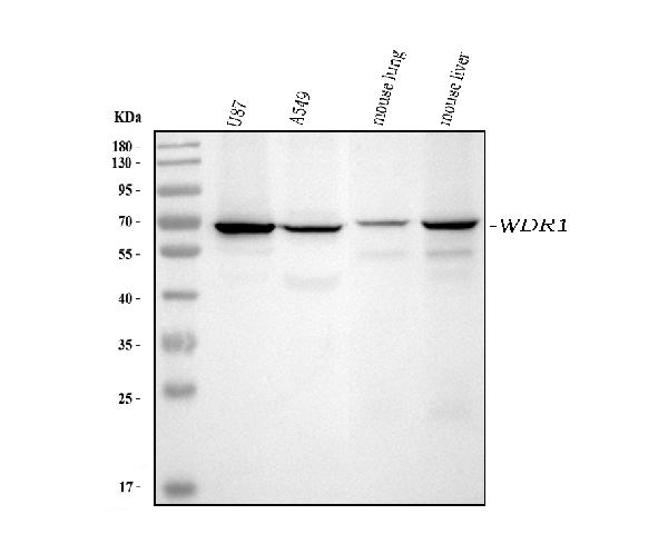
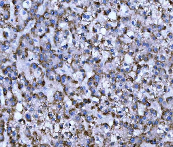
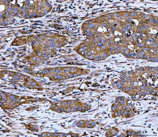
| Description | Anti-WDR1 Antibody Picoband™ (monoclonal, 5C11C8) . Tested in IHC, WB applications. This antibody reacts with Human, Mouse. |
| Reconstitution | Adding 0.2 ml of distilled water will yield a concentration of 500 µg/ml. |
| Gene ID | 9948 |
|---|---|
| Other Names | WD repeat-containing protein 1, Actin-interacting protein 1, AIP1, NORI-1, WDR1 |
| Calculated MW | 66 kDa |
| Application Details | Western blot, 0.25-0.5 µg/ml, Human, Mouse Immunohistochemistry(Paraffin-embedded Section), 2-5 µg/ml, Human |
| Contents | Each vial contains 4 mg Trehalose, 0.9 mg NaCl and 0.2 mg Na2HPO4. |
| Clone Names | Clone: 5C11C8 |
| Immunogen | A synthetic peptide corresponding to a sequence at the C-terminus of human WDR1, different from the related mouse and rat sequences by one amino acid. |
| Purification | Immunogen affinity purified. |
| Storage | At -20°C for one year from date of receipt. After reconstitution, at 4°C for one month. It can also be aliquotted and stored frozen at -20°C for six months. Avoid repeated freezing and thawing. |
| Name | WDR1 |
|---|---|
| Function | Induces disassembly of actin filaments in conjunction with ADF/cofilin family proteins (PubMed:15629458, PubMed:27557945, PubMed:29751004). Enhances cofilin-mediated actin severing (By similarity). Involved in cytokinesis. Involved in chemotactic cell migration by restricting lamellipodial membrane protrusions (PubMed:18494608). Involved in myocardium sarcomere organization. Required for cardiomyocyte growth and maintenance (By similarity). Involved in megakaryocyte maturation and platelet shedding. Required for the establishment of planar cell polarity (PCP) during follicular epithelium development and for cell shape changes during PCP; the function seems to implicate cooperation with CFL1 and/or DSTN/ADF. Involved in the generation/maintenance of cortical tension (By similarity). Involved in assembly and maintenance of epithelial apical cell junctions and plays a role in the organization of the perijunctional actomyosin belt (PubMed:25792565). |
| Cellular Location | Cytoplasm. Cytoplasm, cytoskeleton {ECO:0000250|UniProtKB:Q5RKI0}. Cell projection, podosome. Cell junction |
| Tissue Location | Expressed in peripheral blood mononuclear cells (at protein level). |

Thousands of laboratories across the world have published research that depended on the performance of antibodies from Abcepta to advance their research. Check out links to articles that cite our products in major peer-reviewed journals, organized by research category.
info@abcepta.com, and receive a free "I Love Antibodies" mug.

Provided below are standard protocols that you may find useful for product applications.
Background
WD repeat-containing protein 1 is a protein that in humans is encoded by the WDR1 gene. It is mapped to 4p16.1. This gene encodes a protein containing 9 WD repeats. WD repeats are approximately 30- to 40-amino acid domains containing several conserved residues, mostly including a trp-asp at the C-terminal end. WD domains are involved in protein-protein interactions. The encoded protein may help induce the disassembly of actin filaments. Two transcript variants encoding different isoforms have been found for this gene.
If you have used an Abcepta product and would like to share how it has performed, please click on the "Submit Review" button and provide the requested information. Our staff will examine and post your review and contact you if needed.
If you have any additional inquiries please email technical services at tech@abcepta.com.
Ordering Information
Other Products
Shipping Information
