


 Foundational characteristics of cancer include proliferation, angiogenesis, migration, evasion of apoptosis, and cellular immortality. Find key markers for these cellular processes and antibodies to detect them.
 The SUMOplot™ Analysis Program predicts and scores sumoylation sites in your protein. SUMOylation is a post-translational modification involved in various cellular processes, such as nuclear-cytosolic transport, transcriptional regulation, apoptosis, protein stability, response to stress, and progression through the cell cycle.
 The Autophagy Receptor Motif Plotter predicts and scores autophagy receptor binding sites in your protein. Identifying proteins connected to this pathway is critical to understanding the role of autophagy in physiological as well as pathological processes such as development, differentiation, neurodegenerative diseases, stress, infection, and cancer.
Anti-IFI16 Antibody Picoband™ (monoclonal, 2I3D7)

- SPECIFICATION
- CITATIONS
- PROTOCOLS
- BACKGROUND

Application
| WB, IF, ICC, FC |
|---|---|
| Primary Accession | Q16666 |
| Host | Mouse |
| Isotype | IgG2b |
| Reactivity | Human |
| Clonality | Monoclonal |
| Format | Lyophilized |
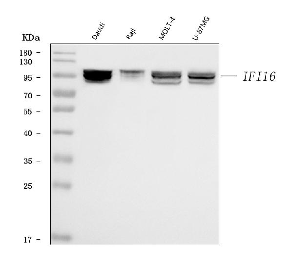
| Description | Anti-IFI16 Antibody Picoband™ (monoclonal, 2I3D7) . Tested in Flow Cytometry, IF, ICC, WB applications. This antibody reacts with Human. |
| Reconstitution | Adding 0.2 ml of distilled water will yield a concentration of 500 µg/ml. |
| Gene ID | 3428 |
|---|---|
| Other Names | Gamma-interferon-inducible protein 16, Ifi-16, Interferon-inducible myeloid differentiation transcriptional activator, IFI16 {ECO:0000303|PubMed:1526658, ECO:0000312|HGNC:HGNC:5395} |
| Calculated MW | 98 kDa |
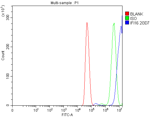
| Application Details | Western blot, 0.25-0.5 µg/ml, Human Immunocytochemistry/Immunofluorescence, 5 µg/ml, Human Flow Cytometry, 1-3 µg/1x10^6 cells, Human |
| Contents | Each vial contains 4 mg Trehalose, 0.9 mg NaCl and 0.2 mg Na2HPO4. |
| Clone Names | Clone: 2I3D7 |
| Immunogen | E.coli-derived human IFI16 recombinant protein (Position: E183-K743). |
| Purification | Immunogen affinity purified. |
| Storage | At -20°C for one year from date of receipt. After reconstitution, at 4°C for one month. It can also be aliquotted and stored frozen at -20°C for six months. Avoid repeated freezing and thawing. |
| Name | IFI16 {ECO:0000303|PubMed:1526658, ECO:0000312|HGNC:HGNC:5395} |
|---|---|
| Function | Binds double-stranded DNA. Binds preferentially to supercoiled DNA and cruciform DNA structures. Seems to be involved in transcriptional regulation. May function as a transcriptional repressor. Could have a role in the regulation of hematopoietic differentiation through activation of unknown target genes. Controls cellular proliferation by modulating the functions of cell cycle regulatory factors including p53/TP53 and the retinoblastoma protein. May be involved in TP53-mediated transcriptional activation by enhancing TP53 sequence-specific DNA binding and modulating TP53 phosphorylation status. Seems to be involved in energy-level-dependent activation of the ATM/ AMPK/TP53 pathway coupled to regulation of autophagy. May be involved in regulation of TP53-mediated cell death also involving BRCA1. May be involved in the senescence of prostate epithelial cells. Involved in innate immune response by recognizing viral dsDNA in the cytosol and probably in the nucleus. After binding to viral DNA in the cytoplasm recruits TMEM173/STING and mediates the induction of IFN-beta. Has anti-inflammatory activity and inhibits the activation of the AIM2 inflammasome, probably via association with AIM2. Proposed to bind viral DNA in the nucleus, such as of Kaposi's sarcoma-associated herpesvirus, and to induce the formation of nuclear caspase-1-activating inflammasome formation via association with PYCARD. Inhibits replication of herpesviruses such as human cytomegalovirus (HCMV) probably by interfering with promoter recruitment of members of the Sp1 family of transcription factors. Necessary to activate the IRF3 signaling cascade during human herpes simplex virus 1 (HHV-1) infection and promotes the assembly of heterochromatin on herpesviral DNA and inhibition of viral immediate- early gene expression and replication. Involved in the MTA1-mediated epigenetic regulation of ESR1 expression in breast cancer. |
| Cellular Location | Nucleus. Cytoplasm. Note=Cellular distribution is dependent on the acetylation status of the multipartite nuclear localization signal (NLS); NLS acetylation promotes cytoplasmic localization Localizes in the nucleus during human herpes simplex virus 1 (HHV-1) infection. |
| Tissue Location | Expressed in peripheral blood leukocytes, fibroblasts and lymphoid cells. Present in myeloid precursors (CD34+) and throughout monocyte development, but its expression is down- regulated in erythroid and polymorphonuclear precursor cells. Present in prostate, ovary and breast (at protein level) |

Thousands of laboratories across the world have published research that depended on the performance of antibodies from Abcepta to advance their research. Check out links to articles that cite our products in major peer-reviewed journals, organized by research category.
info@abcepta.com, and receive a free "I Love Antibodies" mug.

Provided below are standard protocols that you may find useful for product applications.
Background
Gamma-interferon-inducible protein Ifi-16 (Ifi-16) also known as interferon-inducible myeloid differentiation transcriptional activator is a protein that in humans is encoded by the IFI16 gene. This gene encodes a member of the HIN-200 (hematopoietic interferon-inducible nuclear antigens with 200 amino acid repeats) family of cytokines. The encoded protein contains domains involved in DNA binding, transcriptional regulation, and protein-protein interactions. The protein localizes to the nucleoplasm and nucleoli, and interacts with p53 and retinoblastoma-1. It modulates p53 function, and inhibits cell growth in the Ras/Raf signaling pathway. Alternatively spliced transcript variants encoding different isoforms have been found for this gene.
If you have used an Abcepta product and would like to share how it has performed, please click on the "Submit Review" button and provide the requested information. Our staff will examine and post your review and contact you if needed.
If you have any additional inquiries please email technical services at tech@abcepta.com.
Ordering Information
Other Products
Shipping Information
