


 Foundational characteristics of cancer include proliferation, angiogenesis, migration, evasion of apoptosis, and cellular immortality. Find key markers for these cellular processes and antibodies to detect them.
 The SUMOplot™ Analysis Program predicts and scores sumoylation sites in your protein. SUMOylation is a post-translational modification involved in various cellular processes, such as nuclear-cytosolic transport, transcriptional regulation, apoptosis, protein stability, response to stress, and progression through the cell cycle.
 The Autophagy Receptor Motif Plotter predicts and scores autophagy receptor binding sites in your protein. Identifying proteins connected to this pathway is critical to understanding the role of autophagy in physiological as well as pathological processes such as development, differentiation, neurodegenerative diseases, stress, infection, and cancer.
Anti-Golgin 97/GOLGA1 Antibody Picoband™ (monoclonal, 8E4H1)
- SPECIFICATION
- CITATIONS
- PROTOCOLS
- BACKGROUND

Application
| WB, IF, ICC, FC |
|---|---|
| Primary Accession | Q92805 |
| Host | Mouse |
| Isotype | IgG1 |
| Reactivity | Human |
| Clonality | Monoclonal |
| Format | Lyophilized |
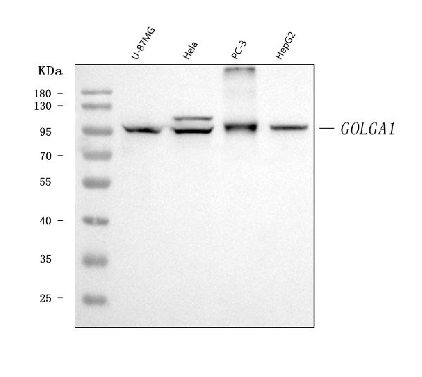
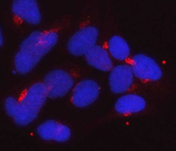
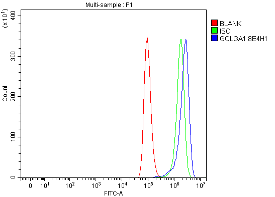
| Description | Anti-Golgin 97/GOLGA1 Antibody Picoband™ (monoclonal, 8E4H1) . Tested in Flow Cytometry, IF, ICC, WB applications. This antibody reacts with Human. |
| Reconstitution | Adding 0.2 ml of distilled water will yield a concentration of 500 µg/ml. |
| Gene ID | 2800 |
|---|---|
| Other Names | Golgin subfamily A member 1, Golgin-97, GOLGA1 |
| Calculated MW | 97 kDa |
| Application Details | Western blot, 0.25-0.5 µg/ml, Human Immunocytochemistry/Immunofluorescence, 5 µg/ml, Human Flow Cytometry, 1-3 µg/1x10^6 cells, Human |
| Contents | Each vial contains 4 mg Trehalose, 0.9 mg NaCl and 0.2 mg Na2HPO4. |
| Clone Names | Clone: 8E4H1 |
| Immunogen | E.coli-derived human Golgin 97/GOLGA1 recombinant protein (Position: M1-K752). |
| Purification | Immunogen affinity purified. |
| Storage | At -20°C for one year from date of receipt. After reconstitution, at 4°C for one month. It can also be aliquotted and stored frozen at -20°C for six months. Avoid repeated freezing and thawing. |
| Name | GOLGA1 |
|---|---|
| Function | Involved in vesicular trafficking at the Golgi apparatus level. Involved in endosome-to-Golgi trafficking. Mechanistically, captures transport vesicles arriving from endosomes via the protein TBC1D23 (PubMed:29084197, PubMed:38552021). Recognized vesicles are then tethered to the trans-Golgi before subsequent SNARE engagement and vesicle fusion. Selectively regulates E-cadherin transport from the trans-Golgi network in tubulovesicular carriers (PubMed:34969853). |
| Cellular Location | Golgi apparatus membrane; Peripheral membrane protein. Golgi apparatus, trans-Golgi network membrane Cytoplasmic vesicle, secretory vesicle, acrosome {ECO:0000250|UniProtKB:Q9CW79} |

Thousands of laboratories across the world have published research that depended on the performance of antibodies from Abcepta to advance their research. Check out links to articles that cite our products in major peer-reviewed journals, organized by research category.
info@abcepta.com, and receive a free "I Love Antibodies" mug.

Provided below are standard protocols that you may find useful for product applications.
Background
Golgin subfamily A member 1 is a protein that in humans is encoded by the GOLGA1 gene. The Golgi apparatus, which participates in glycosylation and transport of proteins and lipids in the secretory pathway, consists of a series of stacked cisternae (flattened membrane sacs). Interactions between the Golgi and microtubules are thought to be important for the reorganization of the Golgi after it fragments during mitosis. This gene encodes one of the golgins, a family of proteins localized to the Golgi. This encoded protein is associated with Sjogren's syndrome.
If you have used an Abcepta product and would like to share how it has performed, please click on the "Submit Review" button and provide the requested information. Our staff will examine and post your review and contact you if needed.
If you have any additional inquiries please email technical services at tech@abcepta.com.
Ordering Information
Other Products
Shipping Information
