


 Foundational characteristics of cancer include proliferation, angiogenesis, migration, evasion of apoptosis, and cellular immortality. Find key markers for these cellular processes and antibodies to detect them.
 The SUMOplot™ Analysis Program predicts and scores sumoylation sites in your protein. SUMOylation is a post-translational modification involved in various cellular processes, such as nuclear-cytosolic transport, transcriptional regulation, apoptosis, protein stability, response to stress, and progression through the cell cycle.
 The Autophagy Receptor Motif Plotter predicts and scores autophagy receptor binding sites in your protein. Identifying proteins connected to this pathway is critical to understanding the role of autophagy in physiological as well as pathological processes such as development, differentiation, neurodegenerative diseases, stress, infection, and cancer.
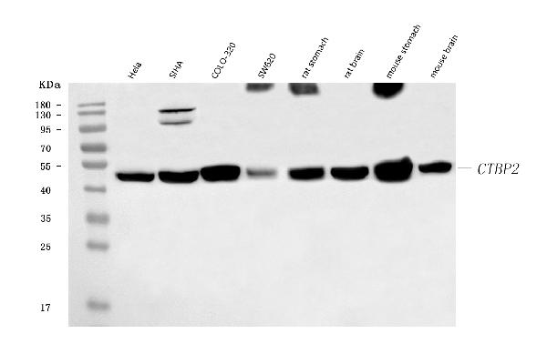
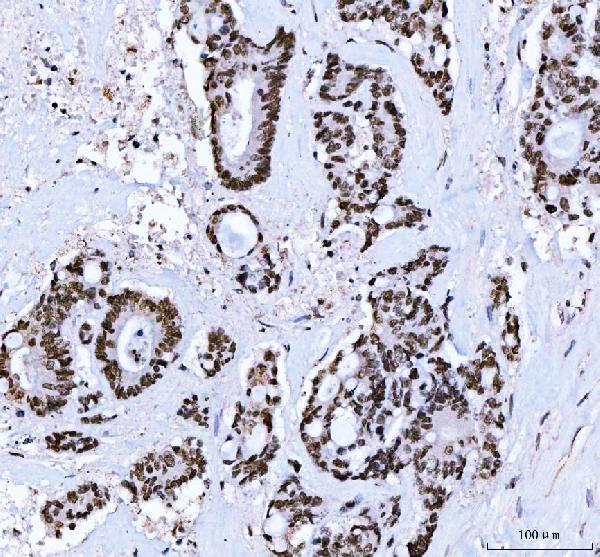
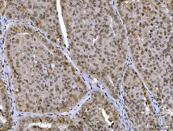
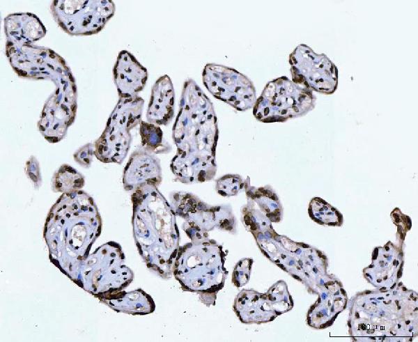
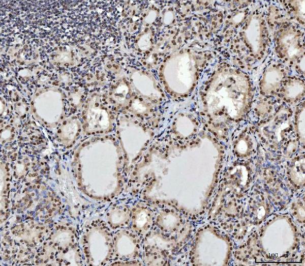
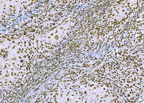
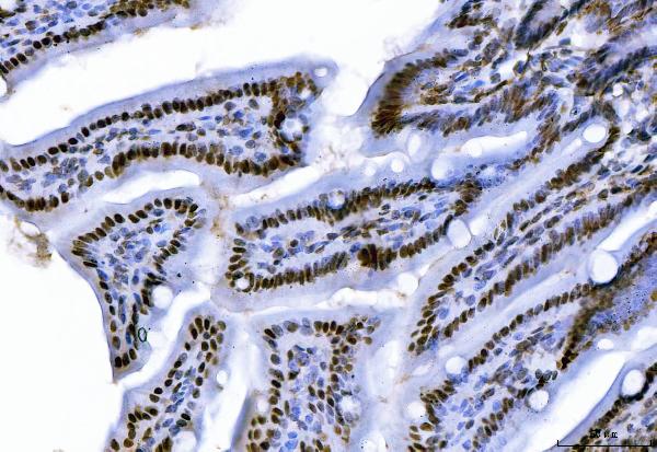
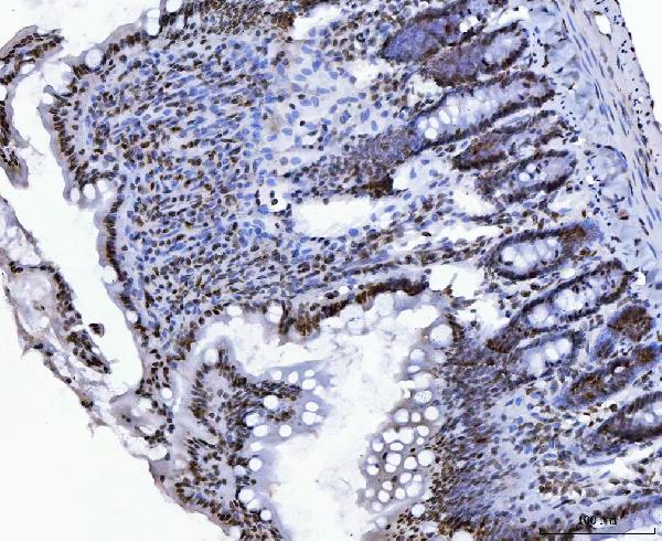
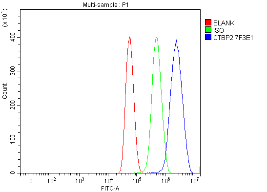
Anti-CTBP2 Antibody Picoband™ (monoclonal, 7F3E1)
- SPECIFICATION
- CITATIONS
- PROTOCOLS
- BACKGROUND

Application
| WB, IHC, FC |
|---|---|
| Primary Accession | P56545 |
| Host | Mouse |
| Isotype | IgG2a |
| Reactivity | Rat, Human, Mouse |
| Clonality | Monoclonal |
| Format | Lyophilized |
| Description | Anti-CTBP2 Antibody Picoband™ (monoclonal, 7F3E1) . Tested in Flow Cytometry, IHC, WB applications. This antibody reacts with Human, Mouse, Rat. |
| Reconstitution | Adding 0.2 ml of distilled water will yield a concentration of 500 µg/ml. |
| Gene ID | 1488 |
|---|---|
| Other Names | C-terminal-binding protein 2, CtBP2, CTBP2 |
| Calculated MW | 49 kDa |
| Application Details | Western blot, 0.25-0.5 µg/ml, Human, Mouse, Rat Immunohistochemistry(Paraffin-embedded Section), 2-5 µg/ml, Human, Mouse, Rat Flow Cytometry, 1-3 µg/1x10^6 cells, Human |
| Contents | Each vial contains 4 mg Trehalose, 0.9 mg NaCl and 0.2 mg Na2HPO4. |
| Clone Names | Clone: 7F3E1 |
| Immunogen | E.coli-derived human CTBP2 recombinant protein (Position: H321-Q445). human CTBP2 shares 99.2% and 98.4% amino acid (aa) sequence identity with mouse and rat CTBP2, respectively. |
| Purification | Immunogen affinity purified. |
| Storage | At -20°C for one year from date of receipt. After reconstitution, at 4°C for one month. It can also be aliquotted and stored frozen at -20°C for six months. Avoid repeated freezing and thawing. |
| Name | CTBP2 |
|---|---|
| Function | Corepressor targeting diverse transcription regulators. Functions in brown adipose tissue (BAT) differentiation (By similarity). |
| Cellular Location | Nucleus. Synapse. |
| Tissue Location | Ubiquitous. Highest levels in heart, skeletal muscle, and pancreas |

Thousands of laboratories across the world have published research that depended on the performance of antibodies from Abcepta to advance their research. Check out links to articles that cite our products in major peer-reviewed journals, organized by research category.
info@abcepta.com, and receive a free "I Love Antibodies" mug.

Provided below are standard protocols that you may find useful for product applications.
Background
The E1a region of group C adenoviruses encodes 2 nearly identical proteins that are largely responsible for the oncogenic properties of adenoviruses. The CTBP1 protein binds to the C-terminal half of these E1A proteins. It's predicted that CTBP2 is a 445-amino acid protein and it is 72% identical to CTBP1. The CTBP2 gene is mapped to chromosome 10q26.13. CTBP2 is a mammalian corepressor that targets diverse transcriptional regulators. It bounds the short medial portion of delta-EF1 containing the PLDLSL motif and it enhances transrepression activity of delta-EF1.
If you have used an Abcepta product and would like to share how it has performed, please click on the "Submit Review" button and provide the requested information. Our staff will examine and post your review and contact you if needed.
If you have any additional inquiries please email technical services at tech@abcepta.com.
Ordering Information
Other Products
Shipping Information
