


 Foundational characteristics of cancer include proliferation, angiogenesis, migration, evasion of apoptosis, and cellular immortality. Find key markers for these cellular processes and antibodies to detect them.
 The SUMOplot™ Analysis Program predicts and scores sumoylation sites in your protein. SUMOylation is a post-translational modification involved in various cellular processes, such as nuclear-cytosolic transport, transcriptional regulation, apoptosis, protein stability, response to stress, and progression through the cell cycle.
 The Autophagy Receptor Motif Plotter predicts and scores autophagy receptor binding sites in your protein. Identifying proteins connected to this pathway is critical to understanding the role of autophagy in physiological as well as pathological processes such as development, differentiation, neurodegenerative diseases, stress, infection, and cancer.
> home > Products > Primary Antibodies > Signal Transduction > Anti-Phospho-AS160 (T642) Rabbit Monoclonal Antibody
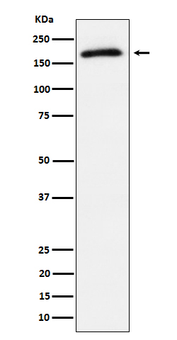
Anti-Phospho-AS160 (T642) Rabbit Monoclonal Antibody
- SPECIFICATION
- CITATIONS
- PROTOCOLS
- BACKGROUND

Application
| WB |
|---|---|
| Primary Accession | O60343 |
| Host | Rabbit |
| Isotype | Rabbit IgG |
| Reactivity | Human |
| Clonality | Monoclonal |
| Format | Liquid |
| Description | Anti-Phospho-AS160 (T642) Rabbit Monoclonal Antibody . Tested in WB applications. This antibody reacts with Human. |
| Gene ID | 9882 |
|---|---|
| Other Names | TBC1 domain family member 4, Akt substrate of 160 kDa, AS160, TBC1D4, AS160, KIAA0603 |
| Calculated MW | 146563 Da |
| Application Details | WB 1:500-1:2000 |
| Contents | Rabbit IgG in phosphate buffered saline, pH 7.4, 150mM NaCl, 0.02% sodium azide and 50% glycerol, 0.4-0.5mg/ml BSA. |
| Clone Names | Clone: 31T47 |
| Immunogen | A synthesized peptide derived from human Phospho-AS160 (T642) |
| Purification | Affinity-chromatography |
| Storage | Store at -20°C for one year. For short term storage and frequent use, store at 4°C for up to one month. Avoid repeated freeze-thaw cycles. |
| Name | TBC1D4 |
|---|---|
| Synonyms | AS160, KIAA0603 |
| Function | May act as a GTPase-activating protein for RAB2A, RAB8A, RAB10 and RAB14. Isoform 2 promotes insulin-induced glucose transporter SLC2A4/GLUT4 translocation at the plasma membrane, thus increasing glucose uptake. |
| Cellular Location | Cytoplasm. Note=Isoform 2 shows a cytoplasmic perinuclear localization in a myoblastic cell line in resting and insulin-stimulated cells |
| Tissue Location | Widely expressed. Isoform 2 is the highest overexpressed in most tissues. Isoform 1 is highly expressed in skeletal muscle and heart, but was not detectable in the liver nor in adipose tissue. Isoform 2 is strongly expressed in adrenal and thyroid gland, and also in lung, kidney, colon, brain and adipose tissue Isoform 2 is moderately expressed in skeletal muscle. Expressed in pancreatic Langerhans islets, including beta cells (at protein level) Expression is decreased by twofold in pancreatic islets in type 2 diabetes patients compared to control subjects. Up-regulated in T-cells from patients with atopic dermatitis. |
Research Areas
Citations (0)

Thousands of laboratories across the world have published research that depended on the performance of antibodies from Abcepta to advance their research. Check out links to articles that cite our products in major peer-reviewed journals, organized by research category.
Submit your citation using an Abcepta antibody to
info@abcepta.com, and receive a free "I Love Antibodies" mug.
info@abcepta.com, and receive a free "I Love Antibodies" mug.

Application Protocols
Provided below are standard protocols that you may find useful for product applications.
Abcepta welcomes feedback from its customers.
If you have used an Abcepta product and would like to share how it has performed, please click on the "Submit Review" button and provide the requested information. Our staff will examine and post your review and contact you if needed.
If you have any additional inquiries please email technical services at tech@abcepta.com.
$ 370.00
Cat# ABO16676
Ordering Information
United States
AlbaniaAustraliaAustriaBelgiumBosnia & HerzegovinaBrazilBulgariaCanadaCentral AmericaChinaCroatiaCyprusCzech RepublicDenmarkEstoniaFinlandFranceGermanyGreeceHong KongHungaryIcelandIndiaIndonesiaIrelandIsraelItalyJapanLatviaLithuaniaLuxembourgMacedoniaMalaysiaMaltaMexicoNetherlandsNew ZealandNorwayPakistanPolandPortugalRomaniaSerbiaSingaporeSlovakiaSloveniaSouth AfricaSouth KoreaSpainSwedenSwitzerlandTaiwanTurkeyUnited KingdomUnited StatesVietnamWorldwideOthers
USA Headquarters
(888) 735-7227 / (858) 622-0099 or (858) 875-1900
Other Products
Shipping Information
Domestic orders (in stock items)
Shipped out the same day. Orders placed after 1 PM (PST) will ship out the next business day.
International orders
Contact your local distributors
