


 Foundational characteristics of cancer include proliferation, angiogenesis, migration, evasion of apoptosis, and cellular immortality. Find key markers for these cellular processes and antibodies to detect them.
 The SUMOplot™ Analysis Program predicts and scores sumoylation sites in your protein. SUMOylation is a post-translational modification involved in various cellular processes, such as nuclear-cytosolic transport, transcriptional regulation, apoptosis, protein stability, response to stress, and progression through the cell cycle.
 The Autophagy Receptor Motif Plotter predicts and scores autophagy receptor binding sites in your protein. Identifying proteins connected to this pathway is critical to understanding the role of autophagy in physiological as well as pathological processes such as development, differentiation, neurodegenerative diseases, stress, infection, and cancer.
> home > Products > Primary Antibodies > Signal Transduction > Anti-Palladin Rabbit Monoclonal Antibody
Anti-Palladin Rabbit Monoclonal Antibody
- SPECIFICATION
- CITATIONS
- PROTOCOLS
- BACKGROUND

Application
| WB |
|---|---|
| Primary Accession | Q8WX93 |
| Host | Rabbit |
| Isotype | Rabbit IgG |
| Reactivity | Human |
| Clonality | Monoclonal |
| Format | Liquid |
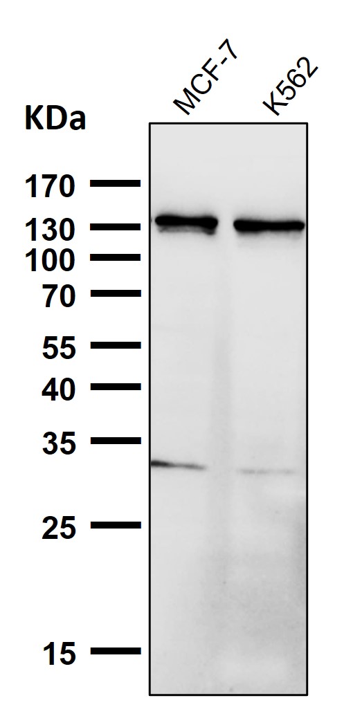
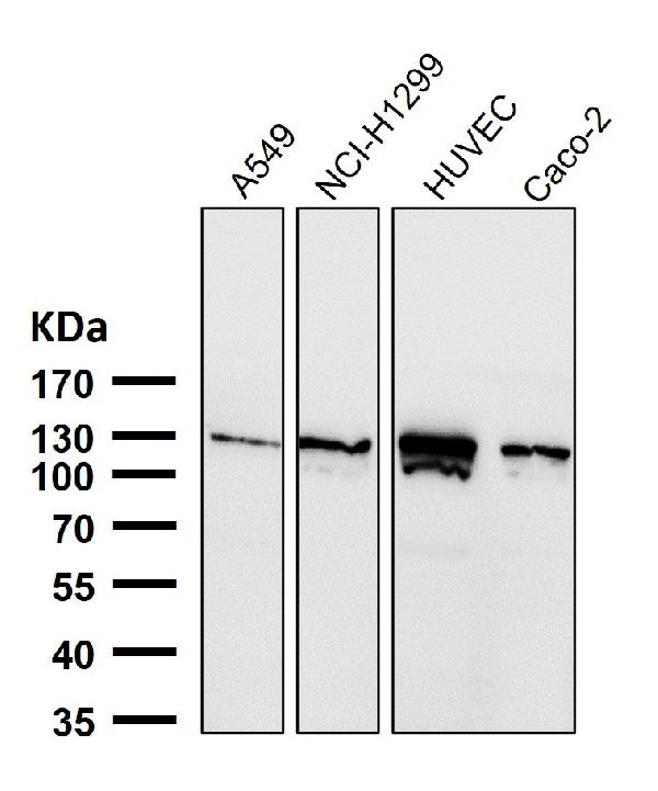
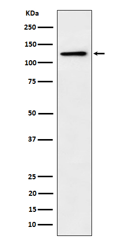
| Description | Anti-Palladin Rabbit Monoclonal Antibody . Tested in WB applications. This antibody reacts with Human. |
| Gene ID | 23022 |
|---|---|
| Other Names | Palladin, SIH002, Sarcoma antigen NY-SAR-77, PALLD, KIAA0992 |
| Calculated MW | 150564 Da |
| Application Details | WB 1:500-1:2000 |
| Contents | Rabbit IgG in phosphate buffered saline, pH 7.4, 150mM NaCl, 0.02% sodium azide and 50% glycerol, 0.4-0.5mg/ml BSA. |
| Clone Names | Clone: 31P55 |
| Immunogen | A synthesized peptide derived from human Palladin |
| Purification | Affinity-chromatography |
| Storage | Store at -20°C for one year. For short term storage and frequent use, store at 4°C for up to one month. Avoid repeated freeze-thaw cycles. |
| Name | PALLD |
|---|---|
| Synonyms | KIAA0992 |
| Function | Cytoskeletal protein required for organization of normal actin cytoskeleton. Roles in establishing cell morphology, motility, cell adhesion and cell-extracellular matrix interactions in a variety of cell types. May function as a scaffolding molecule with the potential to influence both actin polymerization and the assembly of existing actin filaments into higher-order arrays. Binds to proteins that bind to either monomeric or filamentous actin. Localizes at sites where active actin remodeling takes place, such as lamellipodia and membrane ruffles. Different isoforms may have functional differences. Involved in the control of morphological and cytoskeletal changes associated with dendritic cell maturation. Involved in targeting ACTN to specific subcellular foci. |
| Cellular Location | Cytoplasm, cytoskeleton. Cell junction, focal adhesion. Cytoplasm, myofibril, sarcomere, Z line. Cell projection, ruffle. Cell projection, podosome {ECO:0000250|UniProtKB:P0C5E3}. Cell projection, lamellipodium. Cell projection, axon {ECO:0000250|UniProtKB:P0C5E3}. Cell projection, growth cone {ECO:0000250|UniProtKB:P0C5E3}. Note=Localizes to stress fibers and Z lines (PubMed:11598191, PubMed:16125169, PubMed:17322171, PubMed:17537434). Preferentially expressed in the excitatory presynaptic terminals (By similarity). {ECO:0000250|UniProtKB:P0C5E3, ECO:0000269|PubMed:11598191, ECO:0000269|PubMed:16125169, ECO:0000269|PubMed:17322171, ECO:0000269|PubMed:17537434} |
| Tissue Location | Detected in both muscle and non-muscle tissues. High expression in prostate, ovary, colon, and kidney. Not detected in spleen, skeletal muscle, lung and peripheral blood lymphocytes (at protein level). Protein is overexpressed in FA6, HPAF, IMIM-PC2, SUIT-2 and PancTu-II sporadic pancreatic cancer cell lines |
Research Areas
Citations (0)

Thousands of laboratories across the world have published research that depended on the performance of antibodies from Abcepta to advance their research. Check out links to articles that cite our products in major peer-reviewed journals, organized by research category.
Submit your citation using an Abcepta antibody to
info@abcepta.com, and receive a free "I Love Antibodies" mug.
info@abcepta.com, and receive a free "I Love Antibodies" mug.

Application Protocols
Provided below are standard protocols that you may find useful for product applications.
Abcepta welcomes feedback from its customers.
If you have used an Abcepta product and would like to share how it has performed, please click on the "Submit Review" button and provide the requested information. Our staff will examine and post your review and contact you if needed.
If you have any additional inquiries please email technical services at tech@abcepta.com.
$ 370.00
Cat# ABO16684
Ordering Information
United States
AlbaniaAustraliaAustriaBelgiumBosnia & HerzegovinaBrazilBulgariaCanadaCentral AmericaChinaCroatiaCyprusCzech RepublicDenmarkEstoniaFinlandFranceGermanyGreeceHong KongHungaryIcelandIndiaIndonesiaIrelandIsraelItalyJapanLatviaLithuaniaLuxembourgMacedoniaMalaysiaMaltaMexicoNetherlandsNew ZealandNorwayPakistanPolandPortugalRomaniaSerbiaSingaporeSlovakiaSloveniaSouth AfricaSouth KoreaSpainSwedenSwitzerlandTaiwanTurkeyUnited KingdomUnited StatesVietnamWorldwideOthers
USA Headquarters
(888) 735-7227 / (858) 622-0099 or (858) 875-1900
Other Products
Shipping Information
Domestic orders (in stock items)
Shipped out the same day. Orders placed after 1 PM (PST) will ship out the next business day.
International orders
Contact your local distributors
