


 Foundational characteristics of cancer include proliferation, angiogenesis, migration, evasion of apoptosis, and cellular immortality. Find key markers for these cellular processes and antibodies to detect them.
 The SUMOplot™ Analysis Program predicts and scores sumoylation sites in your protein. SUMOylation is a post-translational modification involved in various cellular processes, such as nuclear-cytosolic transport, transcriptional regulation, apoptosis, protein stability, response to stress, and progression through the cell cycle.
 The Autophagy Receptor Motif Plotter predicts and scores autophagy receptor binding sites in your protein. Identifying proteins connected to this pathway is critical to understanding the role of autophagy in physiological as well as pathological processes such as development, differentiation, neurodegenerative diseases, stress, infection, and cancer.
> home > Products > Primary Antibodies > Signal Transduction > Anti-Ceruloplasmin Rabbit Monoclonal Antibody
Anti-Ceruloplasmin Rabbit Monoclonal Antibody
- SPECIFICATION
- CITATIONS
- PROTOCOLS
- BACKGROUND

Application
| WB |
|---|---|
| Primary Accession | P00450 |
| Host | Rabbit |
| Isotype | Rabbit IgG |
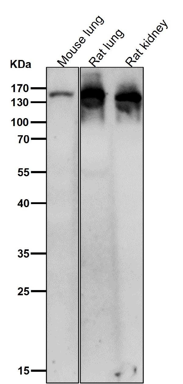
| Reactivity | Rat, Human |
| Clonality | Monoclonal |
| Format | Liquid |
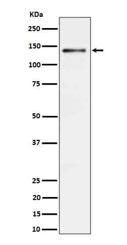
| Description | Anti-Ceruloplasmin Rabbit Monoclonal Antibody . Tested in WB applications. This antibody reacts with Human, Rat. |
| Gene ID | 1356 |
|---|---|
| Other Names | Ceruloplasmin, Cuproxidase ceruloplasmin, 1.16.3.4, Ferroxidase ceruloplasmin, 1.16.3.1, Glutathione peroxidase ceruloplasmin, 1.11.1.9, Glutathione-dependent peroxiredoxin ceruloplasmin, 1.11.1.27, CP (HGNC:2295) |
| Calculated MW | 122219 Da |
| Application Details | WB 1:500-1:2000 |
| Contents | Rabbit IgG in phosphate buffered saline, pH 7.4, 150mM NaCl, 0.02% sodium azide and 50% glycerol, 0.4-0.5mg/ml BSA. |
| Clone Names | Clone: 31C62 |
| Immunogen | A synthesized peptide derived from human Ceruloplasmin |
| Purification | Affinity-chromatography |
| Storage | Store at -20°C for one year. For short term storage and frequent use, store at 4°C for up to one month. Avoid repeated freeze-thaw cycles. |
| Name | CP (HGNC:2295) |
|---|---|
| Function | Multifunctional blue, copper-binding (6-7 atoms per molecule) glycoprotein. It has ferroxidase activity oxidizing Fe(2+) to Fe(3+) without releasing radical oxygen species. It is involved in iron transport across the cell membrane (PubMed:16150804). Copper ions provide a large number of enzymatic activites. Oxidizes highly toxic ferrous ions to the ferric state for further incorporation onto apo- transferrins, catalyzes Cu(+) oxidation and promotes the oxidation of biogenic amines such as norepinephrin and serotonin (PubMed:14623105, PubMed:4643313, PubMed:5912351). Provides Cu(2+) ions for the ascorbate-mediated deaminase degradation of the heparan sulfate chains of GPC1 (By similarity). Has glutathione peroxidase-like activity, can remove both hydrogen peroxide and lipid hydroperoxide in the presence of thiols (PubMed:10481051). Also shows NO-oxidase and NO2 synthase activities that determine endocrine NO homeostasis (PubMed:16906150). |
| Cellular Location | Secreted. Note=Colocalizes with GCP1 in secretory intracellular compartments {ECO:0000250|UniProtKB:P13635} |
| Tissue Location | Expressed by the liver and secreted in plasma. |
Research Areas
Citations (0)

Thousands of laboratories across the world have published research that depended on the performance of antibodies from Abcepta to advance their research. Check out links to articles that cite our products in major peer-reviewed journals, organized by research category.
Submit your citation using an Abcepta antibody to
info@abcepta.com, and receive a free "I Love Antibodies" mug.
info@abcepta.com, and receive a free "I Love Antibodies" mug.

Application Protocols
Provided below are standard protocols that you may find useful for product applications.
Abcepta welcomes feedback from its customers.
If you have used an Abcepta product and would like to share how it has performed, please click on the "Submit Review" button and provide the requested information. Our staff will examine and post your review and contact you if needed.
If you have any additional inquiries please email technical services at tech@abcepta.com.
$ 370.00
Cat# ABO16691
Ordering Information
United States
AlbaniaAustraliaAustriaBelgiumBosnia & HerzegovinaBrazilBulgariaCanadaCentral AmericaChinaCroatiaCyprusCzech RepublicDenmarkEstoniaFinlandFranceGermanyGreeceHong KongHungaryIcelandIndiaIndonesiaIrelandIsraelItalyJapanLatviaLithuaniaLuxembourgMacedoniaMalaysiaMaltaMexicoNetherlandsNew ZealandNorwayPakistanPolandPortugalRomaniaSerbiaSingaporeSlovakiaSloveniaSouth AfricaSouth KoreaSpainSwedenSwitzerlandTaiwanTurkeyUnited KingdomUnited StatesVietnamWorldwideOthers
USA Headquarters
(888) 735-7227 / (858) 622-0099 or (858) 875-1900
Other Products
Shipping Information
Domestic orders (in stock items)
Shipped out the same day. Orders placed after 1 PM (PST) will ship out the next business day.
International orders
Contact your local distributors
