


 Foundational characteristics of cancer include proliferation, angiogenesis, migration, evasion of apoptosis, and cellular immortality. Find key markers for these cellular processes and antibodies to detect them.
 The SUMOplot™ Analysis Program predicts and scores sumoylation sites in your protein. SUMOylation is a post-translational modification involved in various cellular processes, such as nuclear-cytosolic transport, transcriptional regulation, apoptosis, protein stability, response to stress, and progression through the cell cycle.
 The Autophagy Receptor Motif Plotter predicts and scores autophagy receptor binding sites in your protein. Identifying proteins connected to this pathway is critical to understanding the role of autophagy in physiological as well as pathological processes such as development, differentiation, neurodegenerative diseases, stress, infection, and cancer.
> home > Products > Primary Antibodies > Antibody Collections > Pancreas negative > Anti-PICALM Rabbit Monoclonal Antibody
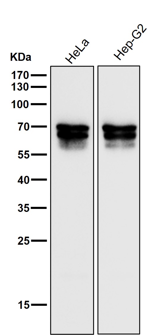
Anti-PICALM Rabbit Monoclonal Antibody
- SPECIFICATION
- CITATIONS
- PROTOCOLS
- BACKGROUND

Application
| WB, IHC |
|---|---|
| Primary Accession | Q13492 |
| Host | Rabbit |
| Isotype | Rabbit IgG |
| Reactivity | Human |
| Clonality | Monoclonal |
| Format | Liquid |
| Description | Anti-PICALM Rabbit Monoclonal Antibody . Tested in WB, IHC, ICC/IF applications. This antibody reacts with Human. |
| Gene ID | 8301 |
|---|---|
| Other Names | Phosphatidylinositol-binding clathrin assembly protein, Clathrin assembly lymphoid myeloid leukemia protein, PICALM, CALM |
| Calculated MW | 70755 Da |
| Application Details | WB 1:500-1:2000 IHC 1:50-1:200 ICC/IF 1:50-1:200 |
| Contents | Rabbit IgG in phosphate buffered saline, pH 7.4, 150mM NaCl, 0.02% sodium azide and 50% glycerol, 0.4-0.5mg/ml BSA. |
| Clone Names | Clone: 32P30 |
| Immunogen | A synthesized peptide derived from human PICALM |
| Purification | Affinity-chromatography |
| Storage | Store at -20°C for one year. For short term storage and frequent use, store at 4°C for up to one month. Avoid repeated freeze-thaw cycles. |
| Name | PICALM |
|---|---|
| Synonyms | CALM |
| Function | Cytoplasmic adapter protein that plays a critical role in clathrin-mediated endocytosis which is important in processes such as internalization of cell receptors, synaptic transmission or removal of apoptotic cells. Recruits AP-2 and attaches clathrin triskelions to the cytoplasmic side of plasma membrane leading to clathrin-coated vesicles (CCVs) assembly (PubMed:10436022, PubMed:16262731, PubMed:27574975). Furthermore, regulates clathrin-coated vesicle size and maturation by directly sensing and driving membrane curvature (PubMed:25898166). In addition to binding to clathrin, mediates the endocytosis of small R- SNARES (Soluble NSF Attachment Protein REceptors) between plasma membranes and endosomes including VAMP2, VAMP3, VAMP4, VAMP7 or VAMP8 (PubMed:21808019, PubMed:22118466, PubMed:23741335). In turn, PICALM- dependent SNARE endocytosis is required for the formation and maturation of autophagic precursors (PubMed:25241929). Modulates thereby autophagy and the turnover of autophagy substrates such as MAPT/TAU or amyloid precursor protein cleaved C-terminal fragment (APP- CTF) (PubMed:24067654, PubMed:25241929). |
| Cellular Location | Cell membrane. Membrane, clathrin-coated pit. Golgi apparatus. Cytoplasmic vesicle, clathrin- coated vesicle. Nucleus. Note=Colocalized with clathrin in the Golgi area (PubMed:10436022). Interaction with PIMREG may target PICALM to the nucleus in some cells (PubMed:16491119) |
| Tissue Location | Expressed in all tissues examined. |
Research Areas
Citations (0)

Thousands of laboratories across the world have published research that depended on the performance of antibodies from Abcepta to advance their research. Check out links to articles that cite our products in major peer-reviewed journals, organized by research category.
Submit your citation using an Abcepta antibody to
info@abcepta.com, and receive a free "I Love Antibodies" mug.
info@abcepta.com, and receive a free "I Love Antibodies" mug.

Application Protocols
Provided below are standard protocols that you may find useful for product applications.
Abcepta welcomes feedback from its customers.
If you have used an Abcepta product and would like to share how it has performed, please click on the "Submit Review" button and provide the requested information. Our staff will examine and post your review and contact you if needed.
If you have any additional inquiries please email technical services at tech@abcepta.com.
$ 370.00
Cat# ABO16759
Ordering Information
United States
AlbaniaAustraliaAustriaBelgiumBosnia & HerzegovinaBrazilBulgariaCanadaCentral AmericaChinaCroatiaCyprusCzech RepublicDenmarkEstoniaFinlandFranceGermanyGreeceHong KongHungaryIcelandIndiaIndonesiaIrelandIsraelItalyJapanLatviaLithuaniaLuxembourgMacedoniaMalaysiaMaltaMexicoNetherlandsNew ZealandNorwayPakistanPolandPortugalRomaniaSerbiaSingaporeSlovakiaSloveniaSouth AfricaSouth KoreaSpainSwedenSwitzerlandTaiwanTurkeyUnited KingdomUnited StatesVietnamWorldwideOthers
USA Headquarters
(888) 735-7227 / (858) 622-0099 or (858) 875-1900
Other Products
Shipping Information
Domestic orders (in stock items)
Shipped out the same day. Orders placed after 1 PM (PST) will ship out the next business day.
International orders
Contact your local distributors
