


 Foundational characteristics of cancer include proliferation, angiogenesis, migration, evasion of apoptosis, and cellular immortality. Find key markers for these cellular processes and antibodies to detect them.
 The SUMOplot™ Analysis Program predicts and scores sumoylation sites in your protein. SUMOylation is a post-translational modification involved in various cellular processes, such as nuclear-cytosolic transport, transcriptional regulation, apoptosis, protein stability, response to stress, and progression through the cell cycle.
 The Autophagy Receptor Motif Plotter predicts and scores autophagy receptor binding sites in your protein. Identifying proteins connected to this pathway is critical to understanding the role of autophagy in physiological as well as pathological processes such as development, differentiation, neurodegenerative diseases, stress, infection, and cancer.
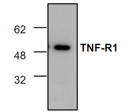
TNF-R1 Antibody
Rabbit Polyclonal Antibody
- SPECIFICATION
- CITATIONS
- PROTOCOLS
- BACKGROUND

Application
| WB, IHC |
|---|---|
| Primary Accession | P25118 |
| Reactivity | Human, Mouse, Rat, Rabbit, Hamster, Monkey, Bovine |
| Host | Rabbit |
| Clonality | Polyclonal |
| Isotype | Rabbit IgG |
| Calculated MW | 50130 Da |
| Gene ID | 21937 |
|---|---|
| Application & Usage | Western blotting (0.5-4 µg/ml) and in Immunohistochemistry (10-20 µg/ml). However, the optimal conditions should be determined individually. The antibody detects cdc42 of human, mouse, rat, and bovine origins. |
| Other Names | TNF-R1, Tumor Necrosis Factor type I, TNFRSF1A, TNFAR, TNF-R55, TNFR60, p55, CD120a |
| Target/Specificity | TNF-RI |
| Antibody Form | Liquid |
| Appearance | Colorless liquid |
| Formulation | 100 µg (0.2 mg/ml) affinity purified rabbit anti-TNF-R1 polyclonal antibody in phosphate buffered saline (PBS), pH 7.2, containing 30% glycerol, 0.5% BSA, 0.01% thimerosal. |
| Handling | The antibody solution should be gently mixed before use. |
| Reconstitution & Storage | -20 °C |
| Background Descriptions | |
| Precautions | TNF-R1 Antibody is for research use only and not for use in diagnostic or therapeutic procedures. |
| Name | Tnfrsf1a |
|---|---|
| Synonyms | Tnfr-1, Tnfr1 |
| Function | Receptor for TNFSF2/TNF-alpha and homotrimeric TNFSF1/lymphotoxin-alpha. The adapter molecule FADD recruits caspase-8 to the activated receptor. The resulting death-inducing signaling complex (DISC) performs caspase-8 proteolytic activation which initiates the subsequent cascade of caspases (aspartate-specific cysteine proteases) mediating apoptosis (By similarity). |
| Cellular Location | Cell membrane; Single-pass type I membrane protein. Golgi apparatus membrane; Single-pass type I membrane protein |

Thousands of laboratories across the world have published research that depended on the performance of antibodies from Abcepta to advance their research. Check out links to articles that cite our products in major peer-reviewed journals, organized by research category.
info@abcepta.com, and receive a free "I Love Antibodies" mug.

Provided below are standard protocols that you may find useful for product applications.
Background
Tumor necrosis factor receptor 1 and 2 (TNF-R1 and TNF-R2) are 55 and 75 kDa. While TNF-R1 and TNF-R2 share 28% sequence homology in the extracellular domains, their intracellular domains lack sequence homology, s µggesting that they differ in their internal signal transduction pathways. TNF-R1 contains an approximately 80 amino acid death domain near its carboxy terminus capable of transmitting an apoptotic signal thro µgh its interaction with TRADD (TNF-R1 associated death domain protein), and subsequent interactions with FADD. TNF-R1 can also activate the transcription factor NFkB via TRAF2 (TNF receptor associated factor 2). The cytoplasmic domain of TNF-R1 can directly interact with Jak kinase, thereby activating the JAK/STAT signal transduction cascade.
If you have used an Abcepta product and would like to share how it has performed, please click on the "Submit Review" button and provide the requested information. Our staff will examine and post your review and contact you if needed.
If you have any additional inquiries please email technical services at tech@abcepta.com.
Ordering Information
Shipping Information
