


 Foundational characteristics of cancer include proliferation, angiogenesis, migration, evasion of apoptosis, and cellular immortality. Find key markers for these cellular processes and antibodies to detect them.
 The SUMOplot™ Analysis Program predicts and scores sumoylation sites in your protein. SUMOylation is a post-translational modification involved in various cellular processes, such as nuclear-cytosolic transport, transcriptional regulation, apoptosis, protein stability, response to stress, and progression through the cell cycle.
 The Autophagy Receptor Motif Plotter predicts and scores autophagy receptor binding sites in your protein. Identifying proteins connected to this pathway is critical to understanding the role of autophagy in physiological as well as pathological processes such as development, differentiation, neurodegenerative diseases, stress, infection, and cancer.
Heme Oxygenase-1 Antibody
Rabbit Polyclonal Antibody
- SPECIFICATION
- CITATIONS
- PROTOCOLS
- BACKGROUND

Application
| WB, IHC, IP |
|---|---|
| Primary Accession | P09601 |
| Reactivity | Human, Mouse, Rat, Rabbit, Hamster, Monkey |
| Host | Rabbit |
| Clonality | Polyclonal |
| Isotype | Rabbit IgG |
| Calculated MW | 32819 Da |
| Gene ID | 3162 |
|---|---|
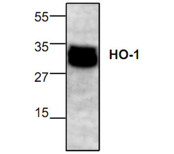
| Application & Usage | Western blotting (0.5-2 µg/ml), immunoprecipitation (10-20 µg/ml), and Immunohistochemistry (10-20 µg/ml). However, the optimal concentrations should be determined individually. The antibody recognizes ~32-35 kDa Heme-Oxygenase-1. Jurkat cell lysate can be used as a positive control. |
| Other Names | HMOX1 , HO , HO-1 , HO1 , OTTHUMP00000028925 , bK286B10 , EC 1.14.99.3 |
| Target/Specificity | Heme Oxygenase-1 |
| Antibody Form | Liquid |
| Appearance | Colorless liquid |
| Formulation | 100 µg (0.5 mg/ml) affinity purified rabbit polyclonal antibody in phosphate-buffered saline (PBS) containing 30% glycerol, 0.5% BSA, and 0.01% thimerosal. |
| Handling | The antibody solution should be gently mixed before use. |
| Reconstitution & Storage | -20 °C |
| Background Descriptions | |
| Precautions | Heme Oxygenase-1 Antibody is for research use only and not for use in diagnostic or therapeutic procedures. |
| Name | HMOX1 |
|---|---|
| Synonyms | HO, HO1 |
| Function | [Heme oxygenase 1]: Catalyzes the oxidative cleavage of heme at the alpha-methene bridge carbon, released as carbon monoxide (CO), to generate biliverdin IXalpha, while releasing the central heme iron chelate as ferrous iron (PubMed:11121422, PubMed:19556236, PubMed:7703255). Affords protection against programmed cell death and this cytoprotective effect relies on its ability to catabolize free heme and prevent it from sensitizing cells to undergo apoptosis (PubMed:20055707). |
| Cellular Location | Endoplasmic reticulum membrane; Single-pass type IV membrane protein; Cytoplasmic side |
| Tissue Location | Expressed at higher levels in renal cancer tissue than in normal tissue (at protein level) |

Thousands of laboratories across the world have published research that depended on the performance of antibodies from Abcepta to advance their research. Check out links to articles that cite our products in major peer-reviewed journals, organized by research category.
info@abcepta.com, and receive a free "I Love Antibodies" mug.

Provided below are standard protocols that you may find useful for product applications.
Background
Heme oxygenase-1 (HO-1) or HSP32 is the inducible isoform of heme oxygenase which catalyzes the NADPH, O2 and cytochrome P450 reductase dependent oxidation of heme to carbon monoxide, iron and biliverdin that is immediately reduced to bilirubin. To date, three heme oxygenase isoforms HO-1, HO-2 and HO-3 have been identified.
If you have used an Abcepta product and would like to share how it has performed, please click on the "Submit Review" button and provide the requested information. Our staff will examine and post your review and contact you if needed.
If you have any additional inquiries please email technical services at tech@abcepta.com.
Ordering Information
Other Products
Shipping Information
