


 Foundational characteristics of cancer include proliferation, angiogenesis, migration, evasion of apoptosis, and cellular immortality. Find key markers for these cellular processes and antibodies to detect them.
 The SUMOplot™ Analysis Program predicts and scores sumoylation sites in your protein. SUMOylation is a post-translational modification involved in various cellular processes, such as nuclear-cytosolic transport, transcriptional regulation, apoptosis, protein stability, response to stress, and progression through the cell cycle.
 The Autophagy Receptor Motif Plotter predicts and scores autophagy receptor binding sites in your protein. Identifying proteins connected to this pathway is critical to understanding the role of autophagy in physiological as well as pathological processes such as development, differentiation, neurodegenerative diseases, stress, infection, and cancer.
Dystrophin Antibody
Rabbit Polyclonal Antibody
- SPECIFICATION
- CITATIONS
- PROTOCOLS
- BACKGROUND

Application
| WB |
|---|---|
| Primary Accession | P11532 |
| Other Accession | NP_004002.2 |
| Reactivity | Human, Mouse, Rat |
| Host | Rabbit |
| Clonality | Polyclonal |
| Isotype | Rabbit IgG |
| Calculated MW | 426750 Da |
| Gene ID | 1756 |
|---|---|
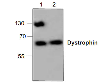
| Application & Usage | Western blotting (0.5-4 µg/ml). However, the optimal conditions should be determined individually. The antibody detects ~60 and 130 kDa of Dystrophin in samples from human, mouse and rat origins. Reactivity to other species has not been determined. |
| Other Names | DMD , DXS272 , DXS269 , DXS230 , Dystrophin, DXS268 , DXS270 , DXS239, DXS142 , CMD3B , DXS206 , DXS164 , BMD |
| Target/Specificity | Dystrophin |
| Antibody Form | Liquid |
| Appearance | Colorless liquid |
| Formulation | 100 µg (0.5 mg/ml) antigen affinity purified rabbit anti-Dystrophin polyclonal antibody in phosphate buffered saline (PBS), pH 7.2, containing 30% glycerol and 0.01% Thimerosal. |
| Handling | The antibody solution should be gently mixed before use. |
| Reconstitution & Storage | -20 °C |
| Background Descriptions | |
| Precautions | Dystrophin Antibody is for research use only and not for use in diagnostic or therapeutic procedures. |
| Name | DMD |
|---|---|
| Function | Anchors the extracellular matrix to the cytoskeleton via F- actin. Ligand for dystroglycan. Component of the dystrophin-associated glycoprotein complex which accumulates at the neuromuscular junction (NMJ) and at a variety of synapses in the peripheral and central nervous systems and has a structural function in stabilizing the sarcolemma. Also implicated in signaling events and synaptic transmission. |
| Cellular Location | Cell membrane, sarcolemma {ECO:0000250|UniProtKB:P11531}; Peripheral membrane protein {ECO:0000250|UniProtKB:P11531}; Cytoplasmic side {ECO:0000250|UniProtKB:P11531}. Cytoplasm, cytoskeleton {ECO:0000250|UniProtKB:P11531}. Postsynaptic cell membrane {ECO:0000250|UniProtKB:P11531}. Note=In muscle cells, sarcolemma localization requires the presence of ANK2, while localization to costameres requires the presence of ANK3. Localizes to neuromuscular junctions (NMJs). In adult muscle, NMJ localization depends upon ANK2 presence, but not in newborn animals. {ECO:0000250|UniProtKB:P11531} |
| Tissue Location | Expressed in muscle fibers accumulating in the costameres of myoplasm at the sarcolemma. Expressed in brain, muscle, kidney, lung and testis. Most tissues contain transcripts of multiple isoforms. Isoform 15: Only isoform to be detected in heart and liver and is also expressed in brain, testis and hepatoma cells |

Thousands of laboratories across the world have published research that depended on the performance of antibodies from Abcepta to advance their research. Check out links to articles that cite our products in major peer-reviewed journals, organized by research category.
info@abcepta.com, and receive a free "I Love Antibodies" mug.

Provided below are standard protocols that you may find useful for product applications.
Background
Dystrophin is one of the actin-binding proteins that are involved in anchoring the cytoskeleton to the plasma membrane. Dystrophin expression is found in muscle brain tissues, where it is located to the inner surface of the plasma membrane. It is s µggested that alternative splicing of the caboxy terminus allows dystrophin to interact with a variety of proteins. Loss of dystrophin-associated proteins in Duchenne afflicted muscle is due to the absence of dystrophin rather than to muscle degradation and lack of dystrophin results in the loss of linkage between the cytoskeleton and extracellular matrix.
If you have used an Abcepta product and would like to share how it has performed, please click on the "Submit Review" button and provide the requested information. Our staff will examine and post your review and contact you if needed.
If you have any additional inquiries please email technical services at tech@abcepta.com.
Ordering Information
Other Products
Shipping Information
