


 Foundational characteristics of cancer include proliferation, angiogenesis, migration, evasion of apoptosis, and cellular immortality. Find key markers for these cellular processes and antibodies to detect them.
 The SUMOplot™ Analysis Program predicts and scores sumoylation sites in your protein. SUMOylation is a post-translational modification involved in various cellular processes, such as nuclear-cytosolic transport, transcriptional regulation, apoptosis, protein stability, response to stress, and progression through the cell cycle.
 The Autophagy Receptor Motif Plotter predicts and scores autophagy receptor binding sites in your protein. Identifying proteins connected to this pathway is critical to understanding the role of autophagy in physiological as well as pathological processes such as development, differentiation, neurodegenerative diseases, stress, infection, and cancer.
ATG9B Antibody (CT)
Rabbit Polyclonal Antibody
- SPECIFICATION
- CITATIONS
- PROTOCOLS
- BACKGROUND

Application
| WB, IF, ICC, E |
|---|---|
| Primary Accession | Q674R7 |
| Reactivity | Human, Mouse, Rat |
| Host | Rabbit |
| Clonality | Polyclonal |
| Isotype | Rabbit IgG1 |
| Calculated MW | 101019 Da |
| Gene ID | 285973 |
|---|---|
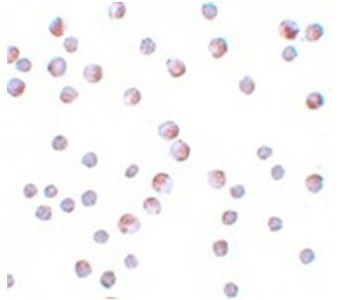
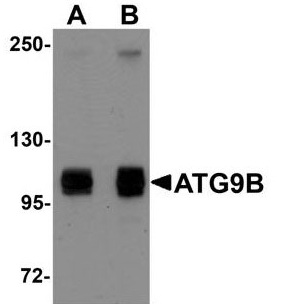
| Positive Control | Western Blot: HeLa cell lysate Immunocytochemistry: HeLa cells Immunoflorescence: HeLa cells |
| Application & Usage | Western Blot: 1 - 2 µg/ml, Immunocytochemistry: 10 µg/ml, Immunoflorescence : 20 µg/ml, ELISA. However, the optimal conditions should be determined individually. |
| Other Names | Autophagy-related protein 9B, APG9L2, APG9-like 2, Nitric oxide synthase 3-overlapping antisense gene protein, NOS3AS |
| Target/Specificity | ATG9B |
| Antibody Form | Liquid |
| Appearance | Colorless liquid |
| Formulation | 100 µg (1 mg/ml) in 1X PBS containing 0.02% sodium azide. |
| Handling | The antibody solution should be gently mixed before use. |
| Reconstitution & Storage | -20 °C |
| Background Descriptions | |
| Precautions | ATG9B Antibody (CT) is for research use only and not for use in diagnostic or therapeutic procedures. |
| Name | ATG9B |
|---|---|
| Function | Phospholipid scramblase involved in autophagy by mediating autophagosomal membrane expansion. Cycles between the preautophagosomal structure/phagophore assembly site (PAS) and the cytoplasmic vesicle pool and supplies membrane for the growing autophagosome. Lipid scramblase activity plays a key role in preautophagosomal structure/phagophore assembly by distributing the phospholipids that arrive through ATG2 (ATG2A or ATG2B) from the cytoplasmic to the luminal leaflet of the bilayer, thereby driving autophagosomal membrane expansion (By similarity). In addition to autophagy, also plays a role in necrotic cell death (By similarity). |
| Cellular Location | Preautophagosomal structure membrane; Multi-pass membrane protein. Note=Under amino acid starvation or rapamycin treatment, redistributes from a juxtanuclear clustered pool to a dispersed peripheral cytosolic pool (PubMed:18936157). The starvation-induced redistribution depends on ULK1 and ATG13 (PubMed:18936157). |
| Tissue Location | Highly expressed in placenta (trophoblast cells) and pituitary gland. Not expressed in vascular endothelial |

Thousands of laboratories across the world have published research that depended on the performance of antibodies from Abcepta to advance their research. Check out links to articles that cite our products in major peer-reviewed journals, organized by research category.
info@abcepta.com, and receive a free "I Love Antibodies" mug.

Provided below are standard protocols that you may find useful for product applications.
Background
Autophagy, the process of bulk degradation of cellular proteins thro µgh an autophagosomic-lysosomal pathway is important for normal growth control and may be defective in tumor cells. It is involved in the preservation of cellular nutrients under starvation conditions as well as the normal turnover of cytosolic components. This process is negatively regulated by TOR (Target of rapamycin) thro µgh phosphorylation of autophagy protein APG1. ATG9B plays a role in autophagy and it's highly expressed in placenta and pituitary gland.
If you have used an Abcepta product and would like to share how it has performed, please click on the "Submit Review" button and provide the requested information. Our staff will examine and post your review and contact you if needed.
If you have any additional inquiries please email technical services at tech@abcepta.com.
Ordering Information
Other Products
Shipping Information
