


 Foundational characteristics of cancer include proliferation, angiogenesis, migration, evasion of apoptosis, and cellular immortality. Find key markers for these cellular processes and antibodies to detect them.
 The SUMOplot™ Analysis Program predicts and scores sumoylation sites in your protein. SUMOylation is a post-translational modification involved in various cellular processes, such as nuclear-cytosolic transport, transcriptional regulation, apoptosis, protein stability, response to stress, and progression through the cell cycle.
 The Autophagy Receptor Motif Plotter predicts and scores autophagy receptor binding sites in your protein. Identifying proteins connected to this pathway is critical to understanding the role of autophagy in physiological as well as pathological processes such as development, differentiation, neurodegenerative diseases, stress, infection, and cancer.
Glutathione Peroxidase 1 Antibody (Clone 2A10)
Mouse Monoclonal Antibody
- SPECIFICATION
- CITATIONS
- PROTOCOLS
- BACKGROUND

Application
| IP |
|---|---|
| Primary Accession | P07203 |
| Reactivity | Human |
| Host | Mouse |
| Clonality | Monoclonal |
| Isotype | Mouse IgG1 |
| Clone Names | Clone 2A10 |
| Calculated MW | 22088 Da |
| Gene ID | 2876 |
|---|---|
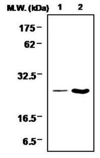
| Positive Control | WB and IP: HL60 cell lysate |
| Application & Usage | IP |
| Other Names | GPX1 |
| Target/Specificity | Glutathione Peroxidase 1 |
| Antibody Form | Liquid |
| Appearance | Colorless liquid |
| Formulation | 100 µl of antibody in HEPES with 0.15 M NaCl, 0.01 % BSA, 0.03 % sodium azide, and 50 % glycerol |
| Handling | The antibody solution should be gently mixed before use. |
| Reconstitution & Storage | -20 °C |
| Background Descriptions | |
| Precautions | Glutathione Peroxidase 1 Antibody (Clone 2A10) is for research use only and not for use in diagnostic or therapeutic procedures. |
| Name | GPX1 (HGNC:4553) |
|---|---|
| Function | Catalyzes the reduction of hydroperoxides in a glutathione- dependent manner thus regulating cellular redox homeostasis (PubMed:11115402, PubMed:36608588). Can reduce small soluble hydroperoxides such as H2O2, cumene hydroperoxide and tert-butyl hydroperoxide, as well as several fatty acid-derived hydroperoxides (PubMed:11115402, PubMed:36608588). In platelets catalyzes the reduction of 12-hydroperoxyeicosatetraenoic acid, the primary product of the arachidonate 12-lipoxygenase pathway (PubMed:11115402). |
| Cellular Location | Cytoplasm {ECO:0000250|UniProtKB:P11352}. Mitochondrion {ECO:0000250|UniProtKB:P11352} |
| Tissue Location | Expressed in platelets (at protein level). |

Thousands of laboratories across the world have published research that depended on the performance of antibodies from Abcepta to advance their research. Check out links to articles that cite our products in major peer-reviewed journals, organized by research category.
info@abcepta.com, and receive a free "I Love Antibodies" mug.

Provided below are standard protocols that you may find useful for product applications.
Background
Glutathione peroxidases (Gpxs) are ubiquitously expressed proteins which catalyze the reduction of hydrogen peroxides and organic hydroperoxides by glutathione. There are several isoforms which differ in their primary structure and localization. The classical cytosolic /mitochondrial GPx1 (cGPx) is a selenium-dependent enzyme, first of the GPx family to be discovered. GPx2, also known as gastrointestinal GPx (GI-GPx), is an intracellular enzyme expressed only at the epithelium of the gastrointestinal tract. Extracellular plasma GPx (pGPx or GPx3) is mainly expressed by the kidney from where it is released into the blood circulation. Phospholipid hydroperoxide GPx4 (PH-GPx) expressed in most tissues, can reduce many hydroperoxides including hydroperoxides integrated in membranes, hydroperoxy lipids in low density lipoprotein or thymine. All mammalian GPx family members, except for the recently described Cys containing GPx3 and epididymis-specific secretory GPx (eGPx or GPx5) isoforms, possess selenocysteine at the active site.
If you have used an Abcepta product and would like to share how it has performed, please click on the "Submit Review" button and provide the requested information. Our staff will examine and post your review and contact you if needed.
If you have any additional inquiries please email technical services at tech@abcepta.com.
Ordering Information
Other Products
Shipping Information
