


 Foundational characteristics of cancer include proliferation, angiogenesis, migration, evasion of apoptosis, and cellular immortality. Find key markers for these cellular processes and antibodies to detect them.
 The SUMOplot™ Analysis Program predicts and scores sumoylation sites in your protein. SUMOylation is a post-translational modification involved in various cellular processes, such as nuclear-cytosolic transport, transcriptional regulation, apoptosis, protein stability, response to stress, and progression through the cell cycle.
 The Autophagy Receptor Motif Plotter predicts and scores autophagy receptor binding sites in your protein. Identifying proteins connected to this pathway is critical to understanding the role of autophagy in physiological as well as pathological processes such as development, differentiation, neurodegenerative diseases, stress, infection, and cancer.
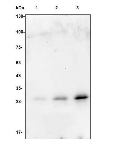
TEV Protease Antibody
Rabbit Polyclonal Antibody
- SPECIFICATION
- CITATIONS
- PROTOCOLS
- BACKGROUND

Application
| WB |
|---|---|
| Reactivity | All Species |
| Host | Rabbit |
| Clonality | Polyclonal |
| Isotype | Rabbit IgG |
| Positive Control | Western Blot: recombinant protein |
|---|---|
| Application & Usage | Western blot: 1-4 µg |
| Other Names | Nuclear inclusion protein A, NIa protein |
| Target/Specificity | TEV protease |
| Antibody Form | Liquid |
| Appearance | Colorless liquid |
| Formulation | 100 µg (0.5 mg/ml) of antibody in PBS pH 7.2, 0.01 % BSA, 0.03 % ProClin®, and 50 % glycerol. |
| Handling | The antibody solution should be gently mixed before use. |
| Reconstitution & Storage | -20 °C |
| Background Descriptions | |
| Precautions | TEV Protease Antibody is for research use only and not for use in diagnostic or therapeutic procedures. |

Thousands of laboratories across the world have published research that depended on the performance of antibodies from Abcepta to advance their research. Check out links to articles that cite our products in major peer-reviewed journals, organized by research category.
info@abcepta.com, and receive a free "I Love Antibodies" mug.

Provided below are standard protocols that you may find useful for product applications.
Background
TEV Protease is a restriction grade protease that has robust activity at 4°C with high specificity and great stability. The optimal temperature for cleavage with this enzyme is 34°C. The protease is used for the removal of affinity tags from fusion proteins. The structure of TEV protease is similar to that of the serine protease family. Like serine proteases, TEV protease utilizes a catalytic triad of residues to hydrolyze peptide bonds. The distinguishing feature of TEV protease, however, is that instead of the serine nucleophile in the triad Ser-Asp-His, there is a cysteine, which may explain the resistance of TEV protease to protease inhibitors which are commonly used.
If you have used an Abcepta product and would like to share how it has performed, please click on the "Submit Review" button and provide the requested information. Our staff will examine and post your review and contact you if needed.
If you have any additional inquiries please email technical services at tech@abcepta.com.
Ordering Information
Shipping Information
