


 Foundational characteristics of cancer include proliferation, angiogenesis, migration, evasion of apoptosis, and cellular immortality. Find key markers for these cellular processes and antibodies to detect them.
 The SUMOplot™ Analysis Program predicts and scores sumoylation sites in your protein. SUMOylation is a post-translational modification involved in various cellular processes, such as nuclear-cytosolic transport, transcriptional regulation, apoptosis, protein stability, response to stress, and progression through the cell cycle.
 The Autophagy Receptor Motif Plotter predicts and scores autophagy receptor binding sites in your protein. Identifying proteins connected to this pathway is critical to understanding the role of autophagy in physiological as well as pathological processes such as development, differentiation, neurodegenerative diseases, stress, infection, and cancer.
CD1e Antibody
Rabbit Polyclonal Antibody
- SPECIFICATION
- CITATIONS
- PROTOCOLS
- BACKGROUND

Application
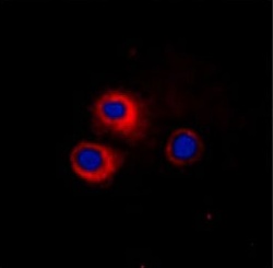
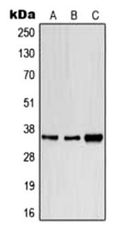
| WB, IF, ICC |
|---|---|
| Primary Accession | P15812 |
| Reactivity | Human, Mouse, Rat |
| Host | Rabbit |
| Clonality | Polyclonal |
| Isotype | Rabbit IgG |
| Calculated MW | 43626 Da |
| Gene ID | 913 |
|---|---|
| Positive Control | WB: HeLa, SP20, H9C2 whole cell lysates, IF/IC: HeLa cells |
| Application & Usage | WB: 1:500 - 1:1000, IF/IC: 1:100 – 1:500 |
| Other Names | T-cell surface glycoprotein CD1e membrane-associated; hCD1e; R2G1; CD1e |
| Target/Specificity | CD1e |
| Antibody Form | Liquid |
| Appearance | Colorless liquid |
| Formulation | 1 mg/ml in 0.42% Potassium phosphate, 0.87% Sodium chloride, pH 7.3, 30% glycerol, and 0.01% sodium azide. |
| Handling | The antibody solution should be gently mixed before use. |
| Reconstitution & Storage | -20 °C |
| Background Descriptions | |
| Precautions | CD1e Antibody is for research use only and not for use in diagnostic or therapeutic procedures. |
| Name | CD1E |
|---|---|
| Function | T-cell surface glycoprotein CD1e, soluble binds diacetylated lipids, including phosphatidyl inositides and diacylated sulfoglycolipids, and is required for the presentation of glycolipid antigens on the cell surface. The membrane-associated form is not active. |
| Cellular Location | [T-cell surface glycoprotein CD1e, membrane- associated]: Golgi apparatus membrane; Single-pass type I membrane protein. Early endosome. Late endosome. Note=Predominantly localized in the trans-Golgi network in immature dendritic cells, and as a cleaved, soluble protein in the lysosome lumen of mature dendritic cells |
| Tissue Location | Expressed on cortical thymocytes, dendritic cells, Langerhans cells, on certain T-cell leukemias, and in various other tissues. |

Thousands of laboratories across the world have published research that depended on the performance of antibodies from Abcepta to advance their research. Check out links to articles that cite our products in major peer-reviewed journals, organized by research category.
info@abcepta.com, and receive a free "I Love Antibodies" mug.

Provided below are standard protocols that you may find useful for product applications.
Background
CD1E encodes a member of the CD1 family of transmembrane glycoproteins, which are structurally related to the major histocompatibility complex (MHC) proteins and form heterodimers with beta-2-microglobulin. The CD1 proteins mediate the presentation of primarily lipid and glycolipid antigens of self or microbial origin to T cells. The human genome contains five CD1 family genes organized in a cluster on chromosome 1. The CD1 family members are thought to differ in their cellular localization and specificity for particular lipid ligands. The protein encoded by this gene localizes within Golgi compartments, endosomes, and lysosomes, and is cleaved into a stable soluble form. The soluble form is required for the intracellular processing of some glycolipids into a form that can be presented by other CD1 family members.
If you have used an Abcepta product and would like to share how it has performed, please click on the "Submit Review" button and provide the requested information. Our staff will examine and post your review and contact you if needed.
If you have any additional inquiries please email technical services at tech@abcepta.com.
Ordering Information
Other Products
Shipping Information
