


 Foundational characteristics of cancer include proliferation, angiogenesis, migration, evasion of apoptosis, and cellular immortality. Find key markers for these cellular processes and antibodies to detect them.
 The SUMOplot™ Analysis Program predicts and scores sumoylation sites in your protein. SUMOylation is a post-translational modification involved in various cellular processes, such as nuclear-cytosolic transport, transcriptional regulation, apoptosis, protein stability, response to stress, and progression through the cell cycle.
 The Autophagy Receptor Motif Plotter predicts and scores autophagy receptor binding sites in your protein. Identifying proteins connected to this pathway is critical to understanding the role of autophagy in physiological as well as pathological processes such as development, differentiation, neurodegenerative diseases, stress, infection, and cancer.
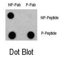
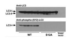
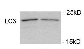
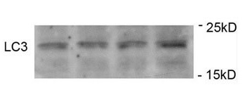
Phospho-LC3C(S12) Antibody
Rabbit Polyclonal Antibody
- SPECIFICATION
- CITATIONS
- PROTOCOLS
- BACKGROUND

Application
| WB, DB |
|---|---|
| Primary Accession | Q9GZQ8 |
| Reactivity | Human, Mouse, Rat, Bovine |
| Host | Rabbit |
| Clonality | Polyclonal |
| Isotype | Rabbit Ig |
| Calculated MW | 14688 Da |
| Gene ID | 81631 |
|---|---|
| Positive Control | WB: SH-SY5Y cells, Dot blot: Phospho and non-phospho peptides |
| Application & Usage | WB: ~1:1000, DB: 1:500. |
| Other Names | MAP1LC3A; Microtubule-associated proteins 1A/1B light chain 3A; Autophagy-related protein LC3 A; Autophagy-related ubiquitin-like modifier LC3 A; MAP1 light chain 3-like protein 1; Microtubule-associated protein 1 light chain 3 alpha |
| Target/Specificity | LC3 |
| Antibody Form | Liquid |
| Appearance | Colorless liquid |
| Formulation | Supplied in PBS with 0.09% (W/V) sodium azide. |
| Handling | The antibody solution should be gently mixed before use. |
| Reconstitution & Storage | -20 °C |
| Background Descriptions | |
| Precautions | Phospho-LC3C(S12) Antibody is for research use only and not for use in diagnostic or therapeutic procedures. |
| Name | MAP1LC3B (HGNC:13352) |
|---|---|
| Synonyms | MAP1ALC3 |
| Function | Ubiquitin-like modifier involved in formation of autophagosomal vacuoles (autophagosomes) (PubMed:20418806, PubMed:23209295, PubMed:28017329). Plays a role in mitophagy which contributes to regulate mitochondrial quantity and quality by eliminating the mitochondria to a basal level to fulfill cellular energy requirements and preventing excess ROS production (PubMed:23209295, PubMed:28017329). In response to cellular stress and upon mitochondria fission, binds C-18 ceramides and anchors autophagolysosomes to outer mitochondrial membranes to eliminate damaged mitochondria (PubMed:22922758). While LC3s are involved in elongation of the phagophore membrane, the GABARAP/GATE-16 subfamily is essential for a later stage in autophagosome maturation (PubMed:20418806, PubMed:23209295, PubMed:28017329). Promotes primary ciliogenesis by removing OFD1 from centriolar satellites via the autophagic pathway (PubMed:24089205). Through its interaction with the reticulophagy receptor TEX264, participates in the remodeling of subdomains of the endoplasmic reticulum into autophagosomes upon nutrient stress, which then fuse with lysosomes for endoplasmic reticulum turnover (PubMed:31006537, PubMed:31006538). Upon nutrient stress, directly recruits cofactor JMY to the phagophore membrane surfaces and promotes JMY's actin nucleation activity and autophagosome biogenesis during autophagy (PubMed:30420355). |
| Cellular Location | Cytoplasmic vesicle, autophagosome membrane; Lipid-anchor Endomembrane system; Lipid-anchor Mitochondrion membrane; Lipid-anchor. Cytoplasm, cytoskeleton {ECO:0000250|UniProtKB:Q9CQV6}. Cytoplasmic vesicle. Note=LC3-II binds to the autophagic membranes. LC3-II localizes with the mitochondrial inner membrane during Parkin-mediated mitophagy (PubMed:28017329). Also localizes to discrete punctae along the ciliary axoneme |
| Tissue Location | Most abundant in heart, brain, skeletal muscle and testis. Little expression observed in liver |

Thousands of laboratories across the world have published research that depended on the performance of antibodies from Abcepta to advance their research. Check out links to articles that cite our products in major peer-reviewed journals, organized by research category.
info@abcepta.com, and receive a free "I Love Antibodies" mug.

Provided below are standard protocols that you may find useful for product applications.
Background
Autophagy is an alternative process of proteasomal degradation for some long-lived proteins or organelles. Alterations in the autophagic-lysosomal compartment have been linked to neuronal death in many neurodegenerative disorders as well as in transmissible neuronal pathologies (prion diseases). Genetic studies in yeast have shown that Autophagy-defective Gene-8 (Atg-8) represents a specific marker for autophagy. Among the four families of mammalian Atg8-related proteins only LC3 (Microtubule-associated Protein1 Light Chain 3) is expressed at sufficient high levels and efficiently recruited to autophagic vesicles in cells and tissues. During autophagy the cytoplasmic form, LC3-I is processed and recruited to autophagosomes, where LC3-II is generated by site specific proteolysis near to the C-terminus. Autophagic vacuoles have been also reported frequently in cardiomyopathies or muscle cells exposed to different experimental settings.
If you have used an Abcepta product and would like to share how it has performed, please click on the "Submit Review" button and provide the requested information. Our staff will examine and post your review and contact you if needed.
If you have any additional inquiries please email technical services at tech@abcepta.com.
Ordering Information
Other Products
Shipping Information
