


 Foundational characteristics of cancer include proliferation, angiogenesis, migration, evasion of apoptosis, and cellular immortality. Find key markers for these cellular processes and antibodies to detect them.
 The SUMOplot™ Analysis Program predicts and scores sumoylation sites in your protein. SUMOylation is a post-translational modification involved in various cellular processes, such as nuclear-cytosolic transport, transcriptional regulation, apoptosis, protein stability, response to stress, and progression through the cell cycle.
 The Autophagy Receptor Motif Plotter predicts and scores autophagy receptor binding sites in your protein. Identifying proteins connected to this pathway is critical to understanding the role of autophagy in physiological as well as pathological processes such as development, differentiation, neurodegenerative diseases, stress, infection, and cancer.
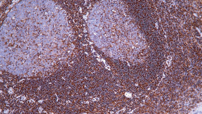
CD44
Mouse Monoclonal antibody(Mab)

- SPECIFICATION
- CITATIONS
- PROTOCOLS
- BACKGROUND

Application
| IHC-P |
|---|---|
| Primary Accession | P16070 |
| Reactivity | Human |
| Host | Mouse |
| Clonality | Monoclonal |
| Clone Names | 344A5C5 |
| Calculated MW | 81538 Da |
| Gene ID | 960 |
|---|---|
| Gene Name | CD44 |
| Other Names | CD44 antigen, CDw44, Epican, Extracellular matrix receptor III, ECMR-III, GP90 lymphocyte homing/adhesion receptor, HUTCH-I, Heparan sulfate proteoglycan, Hermes antigen, Hyaluronate receptor, Phagocytic glycoprotein 1, PGP-1, Phagocytic glycoprotein I, PGP-I, CD44, CD44, LHR, MDU2, MDU3, MIC4 |
| Dilution | IHC-P~~Ready-to-use |
| Storage | Maintain refrigerated at 2-8°C |
| Precautions | CD44 Antibody is for research use only and not for use in diagnostic or therapeutic procedures. |
| Name | CD44 |
|---|---|
| Synonyms | LHR, MDU2, MDU3, MIC4 |
| Function | Receptor for hyaluronic acid (HA). Mediates cell-cell and cell-matrix interactions through its affinity for HA, and possibly also through its affinity for other ligands such as osteopontin, collagens, and matrix metalloproteinases (MMPs). Adhesion with HA plays an important role in cell migration, tumor growth and progression. In cancer cells, may play an important role in invadopodia formation. Also involved in lymphocyte activation, recirculation and homing, and in hematopoiesis. Altered expression or dysfunction causes numerous pathogenic phenotypes. Great protein heterogeneity due to numerous alternative splicing and post-translational modification events. Receptor for LGALS9; the interaction enhances binding of SMAD3 to the FOXP3 promoter, leading to up-regulation of FOXP3 expression and increased induced regulatory T (iTreg) cell stability and suppressive function (By similarity). |
| Cellular Location | Cell membrane {ECO:0000250|UniProtKB:P15379}; Single-pass type I membrane protein {ECO:0000250|UniProtKB:P15379}. Cell projection, microvillus {ECO:0000250|UniProtKB:P15379}. Note=Colocalizes with actin in membrane protrusions at wounding edges. Co-localizes with RDX, EZR and MSN in microvilli. {ECO:0000250|UniProtKB:P15379} |
| Tissue Location | Isoform 10 (epithelial isoform) is expressed by cells of epithelium and highly expressed by carcinomas Expression is repressed in neuroblastoma cells |
Research Areas
Citations (0)

Thousands of laboratories across the world have published research that depended on the performance of antibodies from Abcepta to advance their research. Check out links to articles that cite our products in major peer-reviewed journals, organized by research category.
Submit your citation using an Abcepta antibody to
info@abcepta.com, and receive a free "I Love Antibodies" mug.
info@abcepta.com, and receive a free "I Love Antibodies" mug.

Application Protocols
Provided below are standard protocols that you may find useful for product applications.
Abcepta welcomes feedback from its customers.
If you have used an Abcepta product and would like to share how it has performed, please click on the "Submit Review" button and provide the requested information. Our staff will examine and post your review and contact you if needed.
If you have any additional inquiries please email technical services at tech@abcepta.com.
$ 157.00
$ 106.00
$ 189.00
$ 247.00
Cat# AD80009-010
Ordering Information
United States
AlbaniaAustraliaAustriaBelgiumBosnia & HerzegovinaBrazilBulgariaCanadaCentral AmericaChinaCroatiaCyprusCzech RepublicDenmarkEstoniaFinlandFranceGermanyGreeceHong KongHungaryIcelandIndiaIndonesiaIrelandIsraelItalyJapanLatviaLithuaniaLuxembourgMacedoniaMalaysiaMaltaMexicoNetherlandsNew ZealandNorwayPakistanPolandPortugalRomaniaSerbiaSingaporeSlovakiaSloveniaSouth AfricaSouth KoreaSpainSwedenSwitzerlandTaiwanTurkeyUnited KingdomUnited StatesVietnamWorldwideOthers
USA Headquarters
(888) 735-7227 / (858) 622-0099 or (858) 875-1900
Other Products
Shipping Information
Domestic orders (in stock items)
Shipped out the same day. Orders placed after 1 PM (PST) will ship out the next business day.
International orders
Contact your local distributors
