


 Foundational characteristics of cancer include proliferation, angiogenesis, migration, evasion of apoptosis, and cellular immortality. Find key markers for these cellular processes and antibodies to detect them.
 The SUMOplot™ Analysis Program predicts and scores sumoylation sites in your protein. SUMOylation is a post-translational modification involved in various cellular processes, such as nuclear-cytosolic transport, transcriptional regulation, apoptosis, protein stability, response to stress, and progression through the cell cycle.
 The Autophagy Receptor Motif Plotter predicts and scores autophagy receptor binding sites in your protein. Identifying proteins connected to this pathway is critical to understanding the role of autophagy in physiological as well as pathological processes such as development, differentiation, neurodegenerative diseases, stress, infection, and cancer.
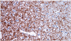
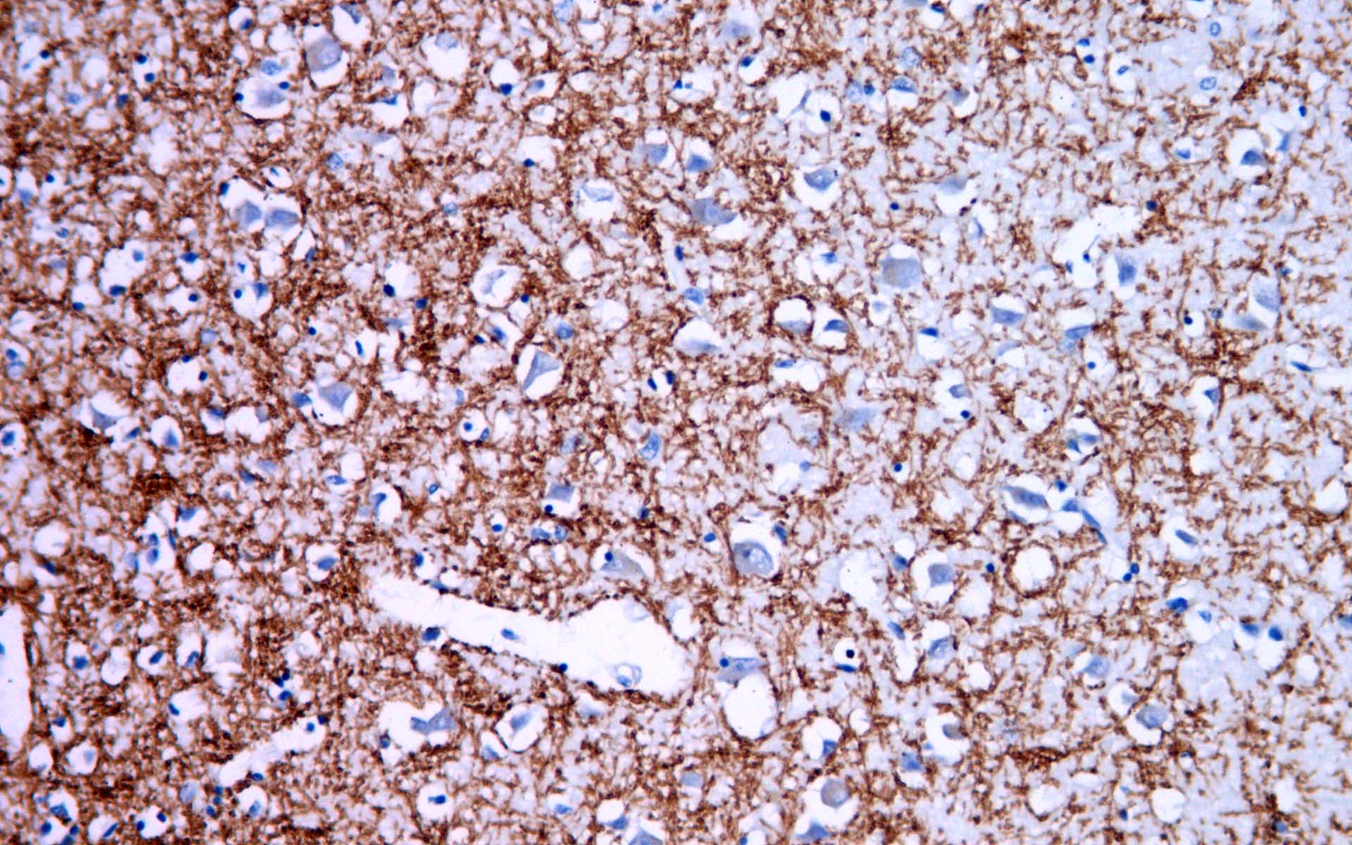
MBP
Rabbit Monoclonal antibody(Mab)
- SPECIFICATION
- CITATIONS
- PROTOCOLS
- BACKGROUND

Application
| IHC-P |
|---|---|
| Primary Accession | P02686 |
| Reactivity | Human |
| Host | Rabbit |
| Clonality | Monoclonal |
| Clone Names | 564B1D5 |
| Calculated MW | 33117 Da |
| Gene ID | 4155 |
|---|---|
| Gene Name | MBP |
| Other Names | Myelin basic protein, MBP, Myelin A1 protein, Myelin membrane encephalitogenic protein, MBP |
| Dilution | IHC-P~~Ready-to-use |
| Storage | Maintain refrigerated at 2-8°C |
| Precautions | Myelin Basic Protein Antibody is for research use only and not for use in diagnostic or therapeutic procedures. |
| Name | MBP |
|---|---|
| Function | The classic group of MBP isoforms (isoform 4-isoform 14) are with PLP the most abundant protein components of the myelin membrane in the CNS. They have a role in both its formation and stabilization. The smaller isoforms might have an important role in remyelination of denuded axons in multiple sclerosis. The non- classic group of MBP isoforms (isoform 1-isoform 3/Golli-MBPs) may preferentially have a role in the early developing brain long before myelination, maybe as components of transcriptional complexes, and may also be involved in signaling pathways in T- cells and neural cells. Differential splicing events combined with optional post-translational modifications give a wide spectrum of isomers, with each of them potentially having a specialized function. Induces T-cell proliferation. |
| Cellular Location | Myelin membrane; Peripheral membrane protein; Cytoplasmic side. Note=Cytoplasmic side of myelin |
| Tissue Location | MBP isoforms are found in both the central and the peripheral nervous system, whereas Golli-MBP isoforms are expressed in fetal thymus, spleen and spinal cord, as well as in cell lines derived from the immune system |
Research Areas
Citations (0)

Thousands of laboratories across the world have published research that depended on the performance of antibodies from Abcepta to advance their research. Check out links to articles that cite our products in major peer-reviewed journals, organized by research category.
Submit your citation using an Abcepta antibody to
info@abcepta.com, and receive a free "I Love Antibodies" mug.
info@abcepta.com, and receive a free "I Love Antibodies" mug.

Application Protocols
Provided below are standard protocols that you may find useful for product applications.
Abcepta welcomes feedback from its customers.
If you have used an Abcepta product and would like to share how it has performed, please click on the "Submit Review" button and provide the requested information. Our staff will examine and post your review and contact you if needed.
If you have any additional inquiries please email technical services at tech@abcepta.com.
$ 127.00
$ 106.00
$ 153.00
$ 182.00
Cat# AD80097-010
Ordering Information
United States
AlbaniaAustraliaAustriaBelgiumBosnia & HerzegovinaBrazilBulgariaCanadaCentral AmericaChinaCroatiaCyprusCzech RepublicDenmarkEstoniaFinlandFranceGermanyGreeceHong KongHungaryIcelandIndiaIndonesiaIrelandIsraelItalyJapanLatviaLithuaniaLuxembourgMacedoniaMalaysiaMaltaMexicoNetherlandsNew ZealandNorwayPakistanPolandPortugalRomaniaSerbiaSingaporeSlovakiaSloveniaSouth AfricaSouth KoreaSpainSwedenSwitzerlandTaiwanTurkeyUnited KingdomUnited StatesVietnamWorldwideOthers
USA Headquarters
(888) 735-7227 / (858) 622-0099 or (858) 875-1900
Other Products
Shipping Information
Domestic orders (in stock items)
Shipped out the same day. Orders placed after 1 PM (PST) will ship out the next business day.
International orders
Contact your local distributors
