


 Foundational characteristics of cancer include proliferation, angiogenesis, migration, evasion of apoptosis, and cellular immortality. Find key markers for these cellular processes and antibodies to detect them.
 The SUMOplot™ Analysis Program predicts and scores sumoylation sites in your protein. SUMOylation is a post-translational modification involved in various cellular processes, such as nuclear-cytosolic transport, transcriptional regulation, apoptosis, protein stability, response to stress, and progression through the cell cycle.
 The Autophagy Receptor Motif Plotter predicts and scores autophagy receptor binding sites in your protein. Identifying proteins connected to this pathway is critical to understanding the role of autophagy in physiological as well as pathological processes such as development, differentiation, neurodegenerative diseases, stress, infection, and cancer.
Functional HMGB1 Antibody, mAb (recombinant)
- SPECIFICATION
- CITATIONS
- PROTOCOLS
- BACKGROUND

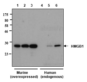
Application
| WB, E |
|---|---|
| Primary Accession | P09429 |
| Reactivity | Human, Mouse, Rat |
| Host | Purified From HEK 293 Cell culture Supernatant. |
| Clonality | Monoclonal |
| Isotype | Human IgG2λ |
| Gene Source | Human |
| Application Note | E,WB(1:1000) |
| Clone Names | Giby-1-4 |
| Calculated MW | 24894 Da |
| Dilution | WB~~1:1000 E~~N/A |
| Description | HMGB1, monoclonal antibody (recombinant) (Giby1-4) is composed of human variable regions (VH and VL) (λ-chain) of immunoglobulin fused to the human lgG2 Fc domain. |
| Gene ID | 3146 |
|---|---|
| Other Names | High Mobility Group Protein B1 |
| Target/Specificity | Recognizes human, mouse and rat HMGB1. |
| Format | Liquid. In PBS containing 10% glycerol and 0.02% sodium azide. |
| Reconstitution & Storage | Stable for at least 1 month after receipt when stored at +4°C. Stable for at least 1 year after receipt when stored at -20°C. |
| Precautions | Functional HMGB1 Antibody, mAb (recombinant) is for research use only and not for use in diagnostic or therapeutic procedures. |
| Name | HMGB1 (HGNC:4983) |
|---|---|
| Synonyms | HMG1 |
| Function | Multifunctional redox sensitive protein with various roles in different cellular compartments. In the nucleus is one of the major chromatin-associated non-histone proteins and acts as a DNA chaperone involved in replication, transcription, chromatin remodeling, V(D)J recombination, DNA repair and genome stability (PubMed:33147444). Proposed to be an universal biosensor for nucleic acids. Promotes host inflammatory response to sterile and infectious signals and is involved in the coordination and integration of innate and adaptive immune responses. In the cytoplasm functions as a sensor and/or chaperone for immunogenic nucleic acids implicating the activation of TLR9-mediated immune responses, and mediates autophagy. Acts as a danger-associated molecular pattern (DAMP) molecule that amplifies immune responses during tissue injury (PubMed:27362237). Released to the extracellular environment can bind DNA, nucleosomes, IL-1 beta, CXCL12, AGER isoform 2/sRAGE, lipopolysaccharide (LPS) and lipoteichoic acid (LTA), and activates cells through engagement of multiple surface receptors (PubMed:34743181). In the extracellular compartment fully reduced HMGB1 (released by necrosis) acts as a chemokine, disulfide HMGB1 (actively secreted) as a cytokine, and sulfonyl HMGB1 (released from apoptotic cells) promotes immunological tolerance (PubMed:23446148, PubMed:23519706, PubMed:23994764, PubMed:25048472). Has proangiogdenic activity (By similarity). May be involved in platelet activation (By similarity). Binds to phosphatidylserine and phosphatidylethanolamide (By similarity). Bound to RAGE mediates signaling for neuronal outgrowth (By similarity). May play a role in accumulation of expanded polyglutamine (polyQ) proteins such as huntingtin (HTT) or TBP (PubMed:23303669, PubMed:25549101). |
| Cellular Location | Nucleus. Chromosome {ECO:0000250|UniProtKB:P10103, ECO:0000250|UniProtKB:P63159, ECO:0000305}. Cytoplasm. Secreted {ECO:0000250|UniProtKB:P63158, ECO:0000269|PubMed:12231511, ECO:0000269|PubMed:14532127, ECO:0000269|PubMed:15944249, ECO:0000269|PubMed:19811284, ECO:0000269|PubMed:22869893, ECO:0000269|PubMed:33147444}. Cell membrane {ECO:0000250|UniProtKB:P63158, ECO:0000250|UniProtKB:P63159, ECO:0000269|PubMed:11154118}; Peripheral membrane protein {ECO:0000250|UniProtKB:P63158, ECO:0000250|UniProtKB:P63159, ECO:0000269|PubMed:11154118}; Extracellular side {ECO:0000250|UniProtKB:P63158, ECO:0000250|UniProtKB:P63159, ECO:0000269|PubMed:11154118}. Endosome {ECO:0000250|UniProtKB:P63158} Endoplasmic reticulum-Golgi intermediate compartment {ECO:0000250|UniProtKB:P63158}. Note=In basal state predominantly nuclear. Shuttles between the cytoplasm and the nucleus (PubMed:12231511, PubMed:17114460). Translocates from the nucleus to the cytoplasm upon autophagy stimulation (PubMed:20819940). Release from macrophages in the extracellular milieu requires the activation of NLRC4 or NLRP3 inflammasomes (By similarity). Passively released to the extracellular milieu from necrotic cells by diffusion, involving the fully reduced HGMB1 which subsequently gets oxidized (PubMed:19811284) Also released from apoptotic cells (PubMed:16855214, PubMed:18631454) Active secretion from a variety of immune and non-immune cells such as macrophages, monocytes, neutrophils, dendritic cells and natural killer cells in response to various stimuli such as LPS and cytokines involves a nonconventional secretory process via secretory lysosomes (PubMed:12231511, PubMed:14532127, PubMed:15944249). Secreted by plasma cells in response to LPS (By similarity). Found on the surface of activated platelets (PubMed:11154118). An increased chromatin association is observed when associated with the adenovirus protein pVII (PubMed:27362237). {ECO:0000250|UniProtKB:P63158, ECO:0000269|PubMed:11154118, ECO:0000269|PubMed:12231511, ECO:0000269|PubMed:14532127, ECO:0000269|PubMed:15944249, ECO:0000269|PubMed:16855214, ECO:0000269|PubMed:17114460, ECO:0000269|PubMed:18631454, ECO:0000269|PubMed:19811284, ECO:0000269|PubMed:20819940, ECO:0000269|PubMed:27362237, ECO:0000305|PubMed:20123072} |
| Tissue Location | Ubiquitous. Expressed in platelets (PubMed:11154118). |

Thousands of laboratories across the world have published research that depended on the performance of antibodies from Abcepta to advance their research. Check out links to articles that cite our products in major peer-reviewed journals, organized by research category.
info@abcepta.com, and receive a free "I Love Antibodies" mug.

Provided below are standard protocols that you may find useful for product applications.
Background
HMGB1 was originally discovered as an essential DNA-binding protein for regulating p53, NF-kappa and other important proteins. It is secreted from activated dentric cells, macrophage and nectrotic cells, and acts as a ligand for RAGE, TLR-2 and TLR-4 expressed on surrounding cells. As a result, HMGB1 activates Rac, CDC42 and NF-kappa inducing localized innate immunity of damaged tissue, tissue regeneration by recruitment of stem cells and hemostasis by induction of tissue factor expression. HMGB1 is also a causative agent of various diseases as it causes localized inflammation such as arteriosclerosis, chronic rheumatoid arthritis and nephritis.
Anti-HMGB1, mAb (recombinant) (Giby-1-4) is an antibody developed by antibody phage display technology using a human naive antibody gene library. These libraries consist of scFv (single chain fragment variable) composed of VH (variable domain of the human immunoglobulin heavy chain) and VL (variable domain of the human immunoglobulin light chain) connected by a polypeptide linker. The antibody fragments are displayed on the surface of filamentous bacteriophage (M13). This scFv was selected by affinity selection on antigen in a process termed panning. Multiple rounds of panning are performed to enrich for antigen-specific scFv-phage. Monoclonal antibodies are subsequently identified by screening after each round of selection. The selected monoclonal scFv is cloned into an appropriate vector containing a Fc portion of interest and then produced in mammalian cells to generate an IgG like scFv-Fc fusion protein.
If you have used an Abcepta product and would like to share how it has performed, please click on the "Submit Review" button and provide the requested information. Our staff will examine and post your review and contact you if needed.
If you have any additional inquiries please email technical services at tech@abcepta.com.
Ordering Information
Other Products
Shipping Information
