


 Foundational characteristics of cancer include proliferation, angiogenesis, migration, evasion of apoptosis, and cellular immortality. Find key markers for these cellular processes and antibodies to detect them.
 The SUMOplot™ Analysis Program predicts and scores sumoylation sites in your protein. SUMOylation is a post-translational modification involved in various cellular processes, such as nuclear-cytosolic transport, transcriptional regulation, apoptosis, protein stability, response to stress, and progression through the cell cycle.
 The Autophagy Receptor Motif Plotter predicts and scores autophagy receptor binding sites in your protein. Identifying proteins connected to this pathway is critical to understanding the role of autophagy in physiological as well as pathological processes such as development, differentiation, neurodegenerative diseases, stress, infection, and cancer.
Goat Anti-BIF-1 / SH3GLB1 Antibody
Peptide-affinity purified goat antibody
- SPECIFICATION
- CITATIONS
- PROTOCOLS
- BACKGROUND

Application
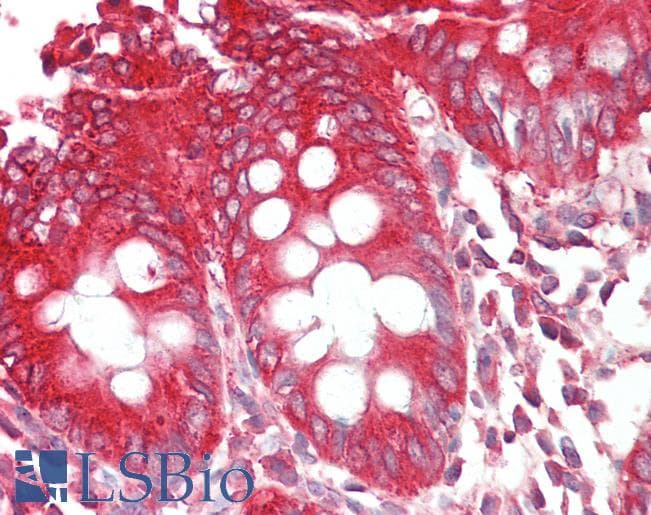
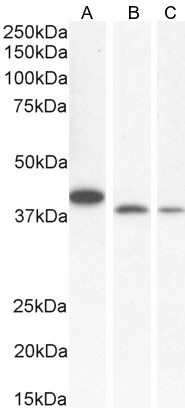
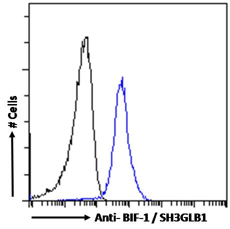
| WB, IHC, FC, Pep-ELISA |
|---|---|
| Primary Accession | Q9Y371 |
| Other Accession | NP_057093, 51100, 54673 (mouse) |
| Reactivity | Human |
| Predicted | Mouse, Rat, Dog |
| Host | Goat |
| Clonality | Polyclonal |
| Concentration | 100ug/200ul |
| Isotype | IgG |
| Calculated MW | 40796 Da |
| Gene ID | 51100 |
|---|---|
| Other Names | Endophilin-B1, Bax-interacting factor 1, Bif-1, SH3 domain-containing GRB2-like protein B1, SH3GLB1, KIAA0491 |
| Dilution | WB~~1:1000 IHC~~1:100~500 FC~~1:10~50 Pep-ELISA~~N/A |
| Format | 0.5 mg IgG/ml in Tris saline (20mM Tris pH7.3, 150mM NaCl), 0.02% sodium azide, with 0.5% bovine serum albumin |
| Storage | Maintain refrigerated at 2-8°C for up to 6 months. For long term storage store at -20°C in small aliquots to prevent freeze-thaw cycles. |
| Precautions | Goat Anti-BIF-1 / SH3GLB1 Antibody is for research use only and not for use in diagnostic or therapeutic procedures. |
| Name | SH3GLB1 |
|---|---|
| Synonyms | KIAA0491 |
| Function | May be required for normal outer mitochondrial membrane dynamics (PubMed:15452144). Required for coatomer-mediated retrograde transport in certain cells (By similarity). May recruit other proteins to membranes with high curvature. May promote membrane fusion (PubMed:11604418). Involved in activation of caspase-dependent apoptosis by promoting BAX/BAK1 activation (PubMed:16227588). Isoform 1 acts proapoptotic in fibroblasts (By similarity). Involved in caspase- independent apoptosis during nutrition starvation and involved in the regulation of autophagy. Activates lipid kinase activity of PIK3C3 during autophagy probably by associating with the PI3K complex II (PI3KC3-C2) (PubMed:17891140). Associated with PI3KC3-C2 during autophagy may regulate the trafficking of ATG9A from the Golgi complex to the peripheral cytoplasm for the formation of autophagosomes by inducing Golgi membrane tubulation and fragmentation (PubMed:21068542). Involved in regulation of degradative endocytic trafficking and cytokinesis, probably in the context of PI3KC3-C2 (PubMed:20643123). Isoform 2 acts antiapoptotic in neuronal cells; involved in maintenance of mitochondrial morphology and promotes neuronal viability (By similarity). |
| Cellular Location | Cytoplasm. Golgi apparatus membrane; Peripheral membrane protein. Mitochondrion outer membrane; Peripheral membrane protein. Cytoplasmic vesicle, autophagosome membrane. Midbody. Note=Association with the Golgi apparatus depends on the cell type (By similarity). Following starvation colocalizes with ATG5 and LC3 autophagy-related protein(s)on autophagosomal membranes (PubMed:17891140). {ECO:0000250, ECO:0000269|PubMed:17891140} |
| Tissue Location | Highly expressed in heart, skeletal muscle, kidney and placenta. Detected at lower levels in brain, colon, thymus, spleen, liver, small intestine, lung and peripheral blood leukocytes |

Thousands of laboratories across the world have published research that depended on the performance of antibodies from Abcepta to advance their research. Check out links to articles that cite our products in major peer-reviewed journals, organized by research category.
info@abcepta.com, and receive a free "I Love Antibodies" mug.

Provided below are standard protocols that you may find useful for product applications.
References
GSK-3beta promotes cell survival by modulating Bif-1-dependent autophagy and cell death. Yang J, et al. J Cell Sci, 2010 Mar 15. PMID 20159967.
Bax activates endophilin B1 oligomerization and lipid membrane vesiculation. Rostovtseva TK, et al. J Biol Chem, 2009 Dec 4. PMID 19805544.
Somatic mutation of pro-cell death Bif-1 gene is rare in common human cancers. Kim MS, et al. APMIS, 2008 Oct. PMID 19132989.
Bif-1 and Bax expression in cutaneous Merkel cell carcinoma. Schlauder SM, et al. J Cutan Pathol, 2009 Jan. PMID 19125733.
Endophilin B1/Bif-1 stimulates BAX activation independently from its capacity to produce large scale membrane morphological rearrangements. Etxebarria A, et al. J Biol Chem, 2009 Feb 13. PMID 19074440.
If you have used an Abcepta product and would like to share how it has performed, please click on the "Submit Review" button and provide the requested information. Our staff will examine and post your review and contact you if needed.
If you have any additional inquiries please email technical services at tech@abcepta.com.
Ordering Information
Other Products
Shipping Information
