


 Foundational characteristics of cancer include proliferation, angiogenesis, migration, evasion of apoptosis, and cellular immortality. Find key markers for these cellular processes and antibodies to detect them.
 The SUMOplot™ Analysis Program predicts and scores sumoylation sites in your protein. SUMOylation is a post-translational modification involved in various cellular processes, such as nuclear-cytosolic transport, transcriptional regulation, apoptosis, protein stability, response to stress, and progression through the cell cycle.
 The Autophagy Receptor Motif Plotter predicts and scores autophagy receptor binding sites in your protein. Identifying proteins connected to this pathway is critical to understanding the role of autophagy in physiological as well as pathological processes such as development, differentiation, neurodegenerative diseases, stress, infection, and cancer.
Goat Anti-FBP17 / FNBP1 Antibody
Peptide-affinity purified goat antibody
- SPECIFICATION
- CITATIONS
- PROTOCOLS
- BACKGROUND

Application
| WB, E |
|---|---|
| Primary Accession | Q96RU3 |
| Other Accession | NP_055848, 23048 |
| Reactivity | Human |
| Predicted | Mouse, Rat, Pig, Dog |
| Host | Goat |
| Clonality | Polyclonal |
| Concentration | 100ug/200ul |
| Isotype | IgG |
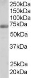
| Calculated MW | 71307 Da |
| Gene ID | 23048 |
|---|---|
| Other Names | Formin-binding protein 1, Formin-binding protein 17, hFBP17, FNBP1, FBP17, KIAA0554 |
| Dilution | WB~~1:1000 E~~N/A |
| Format | 0.5 mg IgG/ml in Tris saline (20mM Tris pH7.3, 150mM NaCl), 0.02% sodium azide, with 0.5% bovine serum albumin |
| Storage | Maintain refrigerated at 2-8°C for up to 6 months. For long term storage store at -20°C in small aliquots to prevent freeze-thaw cycles. |
| Precautions | Goat Anti-FBP17 / FNBP1 Antibody is for research use only and not for use in diagnostic or therapeutic procedures. |
| Name | FNBP1 |
|---|---|
| Synonyms | FBP17, KIAA0554 |
| Function | May act as a link between RND2 signaling and regulation of the actin cytoskeleton (By similarity). Required to coordinate membrane tubulation with reorganization of the actin cytoskeleton during the late stage of clathrin-mediated endocytosis. Binds to lipids such as phosphatidylinositol 4,5-bisphosphate and phosphatidylserine and promotes membrane invagination and the formation of tubules. Also enhances actin polymerization via the recruitment of WASL/N-WASP, which in turn activates the Arp2/3 complex. Actin polymerization may promote the fission of membrane tubules to form endocytic vesicles. May be required for the lysosomal retention of FASLG/FASL. |
| Cellular Location | Cytoplasm. Cytoplasm, cytoskeleton. Cytoplasm, cell cortex. Lysosome. Cytoplasmic vesicle. Cell membrane; Peripheral membrane protein; Cytoplasmic side. Membrane, clathrin-coated pit Note=Enriched in cortical regions coincident with F-actin. Also localizes to endocytic vesicles and lysosomes |
| Tissue Location | Very highly expressed in the epithelial cells of the gastrointestinal tract, respiratory, reproductive and urinary systems. Also highly expressed in brown adipose tissue, cardiomyocytes, enteric ganglia and glucagon producing cells of the pancreas. Expressed in germ cells of the testis and all regions of the brain |

Thousands of laboratories across the world have published research that depended on the performance of antibodies from Abcepta to advance their research. Check out links to articles that cite our products in major peer-reviewed journals, organized by research category.
info@abcepta.com, and receive a free "I Love Antibodies" mug.

Provided below are standard protocols that you may find useful for product applications.
Background
The protein encoded by this gene is a member of the formin-binding-protein family. The protein contains an N-terminal Fer/Cdc42-interacting protein 4 (CIP4) homology (FCH) domain followed by a coiled-coil domain, a proline-rich motif, a second coiled-coil domain, a Rho family protein-binding domain (RBD), and a C-terminal SH3 domain. This protein binds sorting nexin 2 (SNX2), tankyrase (TNKS), and dynamin; an interaction between this protein and formin has not been demonstrated yet in human.
References
Personalized smoking cessation: interactions between nicotine dose, dependence and quit-success genotype score. Rose JE, et al. Mol Med, 2010 Jul-Aug. PMID 20379614.
FBP17 Mediates a Common Molecular Step in the Formation of Podosomes and Phagocytic Cups in Macrophages. Tsuboi S, et al. J Biol Chem, 2009 Mar 27. PMID 19155218.
Identification of interaction partners for individual SH3 domains of Fas ligand associated members of the PCH protein family in T lymphocytes. Linkermann A, et al. Biochim Biophys Acta, 2009 Feb. PMID 19041431.
EFC/F-BAR proteins and the N-WASP-WIP complex induce membrane curvature-dependent actin polymerization. Takano K, et al. EMBO J, 2008 Nov 5. PMID 18923421.
Curved EFC/F-BAR-domain dimers are joined end to end into a filament for membrane invagination in endocytosis. Shimada A, et al. Cell, 2007 May 18. PMID 17512409.
If you have used an Abcepta product and would like to share how it has performed, please click on the "Submit Review" button and provide the requested information. Our staff will examine and post your review and contact you if needed.
If you have any additional inquiries please email technical services at tech@abcepta.com.
Ordering Information
Other Products
Shipping Information
