


 Foundational characteristics of cancer include proliferation, angiogenesis, migration, evasion of apoptosis, and cellular immortality. Find key markers for these cellular processes and antibodies to detect them.
 The SUMOplot™ Analysis Program predicts and scores sumoylation sites in your protein. SUMOylation is a post-translational modification involved in various cellular processes, such as nuclear-cytosolic transport, transcriptional regulation, apoptosis, protein stability, response to stress, and progression through the cell cycle.
 The Autophagy Receptor Motif Plotter predicts and scores autophagy receptor binding sites in your protein. Identifying proteins connected to this pathway is critical to understanding the role of autophagy in physiological as well as pathological processes such as development, differentiation, neurodegenerative diseases, stress, infection, and cancer.
Goat Anti-MBL2 / Mannan-Binding Lectin Antibody
Peptide-affinity purified goat antibody
- SPECIFICATION
- CITATIONS
- PROTOCOLS
- BACKGROUND

Application
| WB, E |
|---|---|
| Primary Accession | P11226 |
| Other Accession | NP_000233, 4153 |
| Reactivity | Human |
| Host | Goat |
| Clonality | Polyclonal |
| Concentration | 100ug/200ul |
| Isotype | IgG |
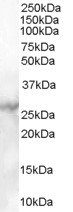
| Calculated MW | 26144 Da |
| Gene ID | 4153 |
|---|---|
| Other Names | Mannose-binding protein C, MBP-C, Collectin-1, MBP1, Mannan-binding protein, Mannose-binding lectin, MBL2, COLEC1, MBL |
| Dilution | WB~~1:1000 E~~N/A |
| Format | 0.5 mg IgG/ml in Tris saline (20mM Tris pH7.3, 150mM NaCl), 0.02% sodium azide, with 0.5% bovine serum albumin |
| Storage | Maintain refrigerated at 2-8°C for up to 6 months. For long term storage store at -20°C in small aliquots to prevent freeze-thaw cycles. |
| Precautions | Goat Anti-MBL2 / Mannan-Binding Lectin Antibody is for research use only and not for use in diagnostic or therapeutic procedures. |
| Name | MBL2 (HGNC:6922) |
|---|---|
| Synonyms | COLEC1, MBL |
| Function | Calcium-dependent lectin involved in innate immune defense (PubMed:35102342). Binds mannose, fucose and N-acetylglucosamine on different microorganisms and activates the lectin complement pathway. Binds to late apoptotic cells, as well as to apoptotic blebs and to necrotic cells, but not to early apoptotic cells, facilitating their uptake by macrophages. May bind DNA. Upon SARS coronavirus-2/SARS-CoV-2 infection, activates the complement lectin pathway which leads to the inhibition SARS-CoV-2 infection and a reduction of the induced inflammatory response (PubMed:35102342). |
| Cellular Location | Secreted. |
| Tissue Location | Plasma protein produced mainly in the liver. |

Thousands of laboratories across the world have published research that depended on the performance of antibodies from Abcepta to advance their research. Check out links to articles that cite our products in major peer-reviewed journals, organized by research category.
info@abcepta.com, and receive a free "I Love Antibodies" mug.

Provided below are standard protocols that you may find useful for product applications.
Background
This gene encodes the soluble mannose-binding lectin or mannose-binding protein found in serum. The protein encoded belongs to the collectin family and is an important element in the innate immune system. The protein recognizes mannose and N-acetylglucosamine on many microorganisms, and is capable of activating the classical complement pathway. Deficiencies of this gene have been associated with susceptibility to autoimmune and infectious diseases.
References
Gene-gene interaction between tuberculosis candidate genes in a South African population. de Wit E, et al. Mamm Genome, 2010 Aug 27. PMID 20799037.
Role of mannose-binding lectin in nosocomial sepsis in critically ill neonates. Auriti C, et al. Hum Immunol, 2010 Aug 21. PMID 20732365.
High frequency of variant alleles of the mannose-binding lectin 2 (MBL2) gene are associated with patients infected by hepatitis B virus. Filho RM, et al. Viral Immunol, 2010 Aug. PMID 20712490.
Association between mannose-binding lectin gene polymorphism and pediatric cytomegalovirus infection. Hu Y, et al. Viral Immunol, 2010 Aug. PMID 20712489.
Mannose-binding lectin geno- and phenotype in patients with type 2 diabetes and myocardial infarction -a report from the DIGAMI 2 trial. Mellbin LG, et al. Diabetes Care, 2010 Aug 6. PMID 20693349.
If you have used an Abcepta product and would like to share how it has performed, please click on the "Submit Review" button and provide the requested information. Our staff will examine and post your review and contact you if needed.
If you have any additional inquiries please email technical services at tech@abcepta.com.
Ordering Information
Other Products
Shipping Information
