


 Foundational characteristics of cancer include proliferation, angiogenesis, migration, evasion of apoptosis, and cellular immortality. Find key markers for these cellular processes and antibodies to detect them.
 The SUMOplot™ Analysis Program predicts and scores sumoylation sites in your protein. SUMOylation is a post-translational modification involved in various cellular processes, such as nuclear-cytosolic transport, transcriptional regulation, apoptosis, protein stability, response to stress, and progression through the cell cycle.
 The Autophagy Receptor Motif Plotter predicts and scores autophagy receptor binding sites in your protein. Identifying proteins connected to this pathway is critical to understanding the role of autophagy in physiological as well as pathological processes such as development, differentiation, neurodegenerative diseases, stress, infection, and cancer.
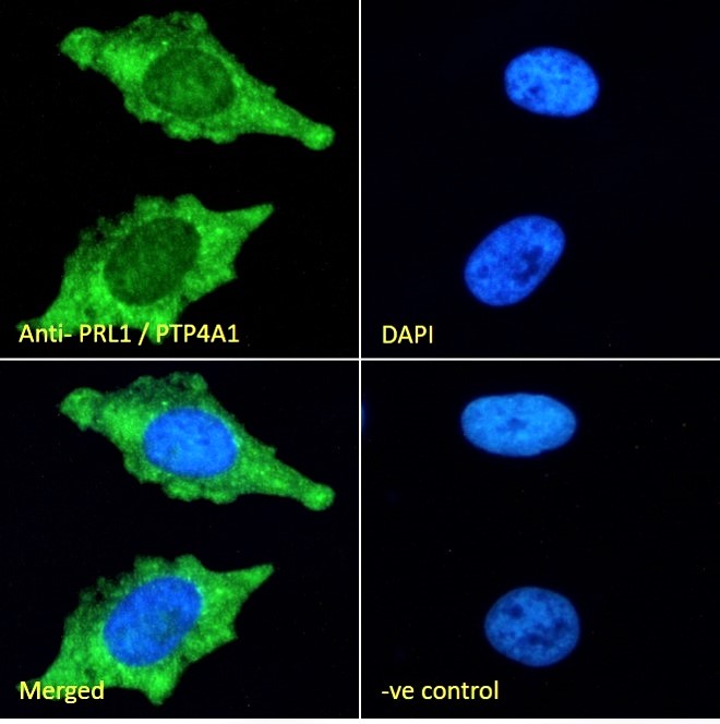
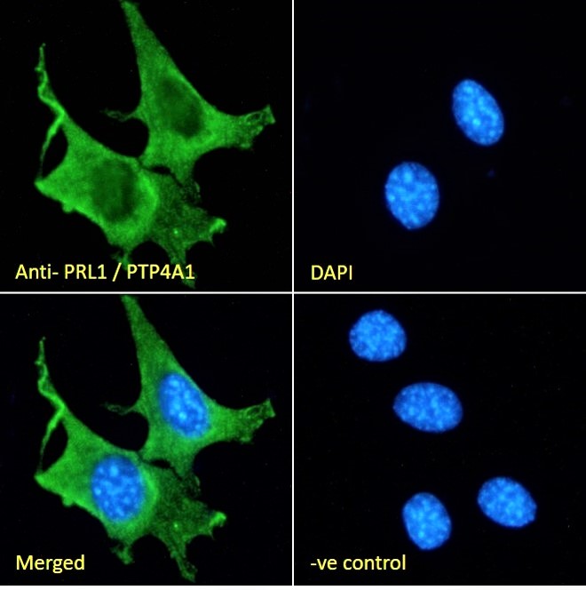
PRL1 / PTP4A1 Antibody (C-Term)
Peptide-affinity purified goat antibody
- SPECIFICATION
- CITATIONS
- PROTOCOLS
- BACKGROUND

Application
| WB, E |
|---|---|
| Primary Accession | Q93096 |
| Other Accession | NP_003454.1, 7803 |
| Reactivity | Human |
| Predicted | Mouse, Rat, Dog |
| Host | Goat |
| Clonality | Polyclonal |
| Concentration | 0.5 mg/ml |
| Isotype | IgG |
| Calculated MW | 19815 Da |
| Gene ID | 7803 |
|---|---|
| Other Names | Protein tyrosine phosphatase type IVA 1, 3.1.3.48, PTP(CAAXI), Protein-tyrosine phosphatase 4a1, Protein-tyrosine phosphatase of regenerating liver 1, PRL-1, PTP4A1, PRL1, PTPCAAX1 |
| Dilution | WB~~1:1000 E~~N/A |
| Format | 0.5 mg/ml in Tris saline, 0.02% sodium azide, pH7.3 with 0.5% bovine serum albumin |
| Storage | Maintain refrigerated at 2-8°C for up to 6 months. For long term storage store at -20°C in small aliquots to prevent freeze-thaw cycles. |
| Precautions | PRL1 / PTP4A1 Antibody (C-Term) is for research use only and not for use in diagnostic or therapeutic procedures. |
| Name | PTP4A1 |
|---|---|
| Synonyms | PRL1, PTPCAAX1 |
| Function | Protein tyrosine phosphatase which stimulates progression from G1 into S phase during mitosis. May play a role in the development and maintenance of differentiating epithelial tissues. Enhances cell proliferation, cell motility and invasive activity, and promotes cancer metastasis. |
| Cellular Location | Cell membrane; Lipid-anchor. Early endosome. Endoplasmic reticulum. Cytoplasm Cytoplasm, cytoskeleton, spindle. Nucleus {ECO:0000250|UniProtKB:Q78EG7}. Note=And mitotic spindle |
| Tissue Location | Expressed in bone marrow, lymph nodes, T lymphocytes, spleen, thymus and tonsil. Overexpressed in tumor cell lines. |

Thousands of laboratories across the world have published research that depended on the performance of antibodies from Abcepta to advance their research. Check out links to articles that cite our products in major peer-reviewed journals, organized by research category.
info@abcepta.com, and receive a free "I Love Antibodies" mug.

Provided below are standard protocols that you may find useful for product applications.
References
The gene encoding human nuclear protein tyrosine phosphatase, PRL-1. Cloning, chromosomal localization, and identification of an intron enhancer. Peng Y, Genin A, Spinner NB, Diamond RH, Taub R. J Biol Chem. 1998 Jul 3;273(27):17286-95. PMID: 9642300
If you have used an Abcepta product and would like to share how it has performed, please click on the "Submit Review" button and provide the requested information. Our staff will examine and post your review and contact you if needed.
If you have any additional inquiries please email technical services at tech@abcepta.com.
Ordering Information
Other Products
Shipping Information
