


 Foundational characteristics of cancer include proliferation, angiogenesis, migration, evasion of apoptosis, and cellular immortality. Find key markers for these cellular processes and antibodies to detect them.
 The SUMOplot™ Analysis Program predicts and scores sumoylation sites in your protein. SUMOylation is a post-translational modification involved in various cellular processes, such as nuclear-cytosolic transport, transcriptional regulation, apoptosis, protein stability, response to stress, and progression through the cell cycle.
 The Autophagy Receptor Motif Plotter predicts and scores autophagy receptor binding sites in your protein. Identifying proteins connected to this pathway is critical to understanding the role of autophagy in physiological as well as pathological processes such as development, differentiation, neurodegenerative diseases, stress, infection, and cancer.
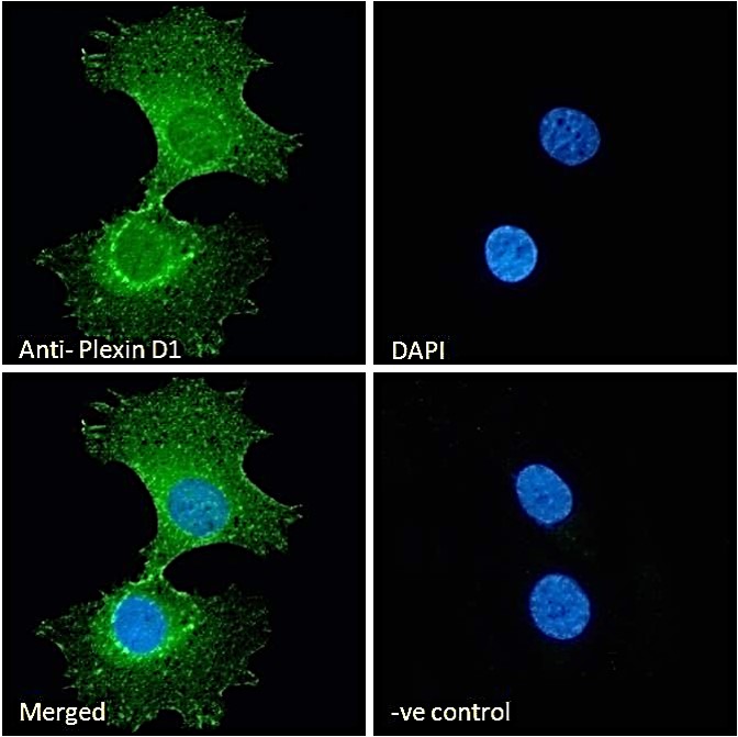
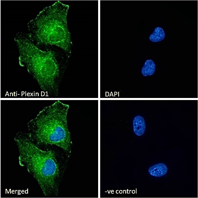
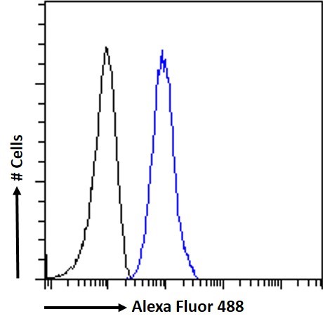
Plexin D1 Antibody (internal region)
Peptide-affinity purified goat antibody
- SPECIFICATION
- CITATIONS
- PROTOCOLS
- BACKGROUND

Application
| IF, FC, Pep-ELISA |
|---|---|
| Primary Accession | Q9Y4D7 |
| Other Accession | NP_055918.2, 23129 |
| Reactivity | Human, Mouse |
| Predicted | Pig, Dog |
| Host | Goat |
| Clonality | Polyclonal |
| Concentration | 0.5 mg/ml |
| Isotype | IgG |
| Calculated MW | 212007 Da |
| Gene ID | 23129 |
|---|---|
| Other Names | Plexin-D1, PLXND1, KIAA0620 |
| Dilution | IF~~1:50~200 FC~~1:10~50 Pep-ELISA~~N/A |
| Format | 0.5 mg/ml in Tris saline, 0.02% sodium azide, pH7.3 with 0.5% bovine serum albumin |
| Storage | Maintain refrigerated at 2-8°C for up to 6 months. For long term storage store at -20°C in small aliquots to prevent freeze-thaw cycles. |
| Precautions | Plexin D1 Antibody (internal region) is for research use only and not for use in diagnostic or therapeutic procedures. |
| Name | PLXND1 |
|---|---|
| Synonyms | KIAA0620 |
| Function | Cell surface receptor for SEMA4A and for class 3 semaphorins, such as SEMA3A, SEMA3C and SEMA3E. Plays an important role in cell-cell signaling, and in regulating the migration of a wide spectrum of cell types. Regulates the migration of thymocytes in the medulla. Regulates endothelial cell migration. Plays an important role in ensuring the specificity of synapse formation. Required for normal development of the heart and vasculature (By similarity). Mediates anti-angiogenic signaling in response to SEMA3E. |
| Cellular Location | Cell membrane {ECO:0000250|UniProtKB:Q3UH93}; Single-pass membrane protein {ECO:0000250|UniProtKB:Q3UH93}. Cell projection, lamellipodium membrane |
| Tissue Location | Detected at low levels in heart, placenta, lung, skeletal muscle, kidney, thymus and liver. Detected at very low levels in brain, colon, spleen, small intestine and peripheral blood leukocytes. |

Thousands of laboratories across the world have published research that depended on the performance of antibodies from Abcepta to advance their research. Check out links to articles that cite our products in major peer-reviewed journals, organized by research category.
info@abcepta.com, and receive a free "I Love Antibodies" mug.

Provided below are standard protocols that you may find useful for product applications.
References
Semaphorin 3E and plexin-D1 control vascular pattern independently of neuropilins. Gu C, Yoshida Y, Livet J, Reimert DV, Mann F, Merte J, Henderson CE, Jessell TM, Kolodkin AL, Ginty DD. Science. 2005 Jan 14;307(5707):265-8. Epub 2004 Nov 18. PMID: 15550623
If you have used an Abcepta product and would like to share how it has performed, please click on the "Submit Review" button and provide the requested information. Our staff will examine and post your review and contact you if needed.
If you have any additional inquiries please email technical services at tech@abcepta.com.
Ordering Information
Other Products
Shipping Information
