


 Foundational characteristics of cancer include proliferation, angiogenesis, migration, evasion of apoptosis, and cellular immortality. Find key markers for these cellular processes and antibodies to detect them.
 The SUMOplot™ Analysis Program predicts and scores sumoylation sites in your protein. SUMOylation is a post-translational modification involved in various cellular processes, such as nuclear-cytosolic transport, transcriptional regulation, apoptosis, protein stability, response to stress, and progression through the cell cycle.
 The Autophagy Receptor Motif Plotter predicts and scores autophagy receptor binding sites in your protein. Identifying proteins connected to this pathway is critical to understanding the role of autophagy in physiological as well as pathological processes such as development, differentiation, neurodegenerative diseases, stress, infection, and cancer.
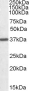
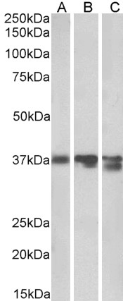
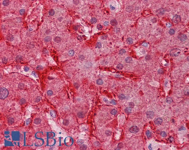
Arginase I Antibody (C-Term)
Peptide-affinity purified goat antibody
- SPECIFICATION
- CITATIONS
- PROTOCOLS
- BACKGROUND

Application
| WB, IHC, Pep-ELISA |
|---|---|
| Primary Accession | P05089 |
| Other Accession | NP_000036.2, 383, 11846 (mouse), 29221 (rat) |
| Reactivity | Human, Mouse, Rat, Pig |
| Predicted | Dog |
| Host | Goat |
| Clonality | Polyclonal |
| Concentration | 0.5 mg/ml |
| Isotype | IgG |
| Calculated MW | 34735 Da |
| Gene ID | 383 |
|---|---|
| Other Names | Arginase-1, 3.5.3.1, Liver-type arginase, Type I arginase, ARG1 |
| Dilution | WB~~1:1000 IHC~~1:100~500 Pep-ELISA~~N/A |
| Format | 0.5 mg/ml in Tris saline, 0.02% sodium azide, pH7.3 with 0.5% bovine serum albumin |
| Storage | Maintain refrigerated at 2-8°C for up to 6 months. For long term storage store at -20°C in small aliquots to prevent freeze-thaw cycles. |
| Precautions | Arginase I Antibody (C-Term) is for research use only and not for use in diagnostic or therapeutic procedures. |
| Name | ARG1 |
|---|---|
| Function | Key element of the urea cycle converting L-arginine to urea and L-ornithine, which is further metabolized into metabolites proline and polyamides that drive collagen synthesis and bioenergetic pathways critical for cell proliferation, respectively; the urea cycle takes place primarily in the liver and, to a lesser extent, in the kidneys. |
| Cellular Location | Cytoplasm. Cytoplasmic granule. Note=Localized in azurophil granules of neutrophils (PubMed:15546957) |
| Tissue Location | Within the immune system initially reported to be selectively expressed in granulocytes (polymorphonuclear leukocytes [PMNs]) (PubMed:15546957). Also detected in macrophages mycobacterial granulomas (PubMed:23749634). Expressed in group2 innate lymphoid cells (ILC2s) during lung disease (PubMed:27043409) |

Thousands of laboratories across the world have published research that depended on the performance of antibodies from Abcepta to advance their research. Check out links to articles that cite our products in major peer-reviewed journals, organized by research category.
info@abcepta.com, and receive a free "I Love Antibodies" mug.

Provided below are standard protocols that you may find useful for product applications.
References
A review of asthma genetics: gene expression studies and recent candidates. Malerba G, Pignatti PF. J Appl Genet. 2005;46(1):93-104. Review. PMID: 15741670
If you have used an Abcepta product and would like to share how it has performed, please click on the "Submit Review" button and provide the requested information. Our staff will examine and post your review and contact you if needed.
If you have any additional inquiries please email technical services at tech@abcepta.com.
Ordering Information
Other Products
Shipping Information
