


 Foundational characteristics of cancer include proliferation, angiogenesis, migration, evasion of apoptosis, and cellular immortality. Find key markers for these cellular processes and antibodies to detect them.
 The SUMOplot™ Analysis Program predicts and scores sumoylation sites in your protein. SUMOylation is a post-translational modification involved in various cellular processes, such as nuclear-cytosolic transport, transcriptional regulation, apoptosis, protein stability, response to stress, and progression through the cell cycle.
 The Autophagy Receptor Motif Plotter predicts and scores autophagy receptor binding sites in your protein. Identifying proteins connected to this pathway is critical to understanding the role of autophagy in physiological as well as pathological processes such as development, differentiation, neurodegenerative diseases, stress, infection, and cancer.
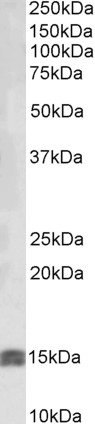
IGF1 Antibody (internal region)
Peptide-affinity purified goat antibody
- SPECIFICATION
- CITATIONS
- PROTOCOLS
- BACKGROUND

Application
| WB, E |
|---|---|
| Primary Accession | P05019 |
| Other Accession | NP_001104753.1, NP_001104754.1, NP_001104755.1, NP_000609.1, 3479, 16000 (mouse), 24482 (rat) |
| Reactivity | Human |
| Predicted | Mouse, Rat, Rabbit, Pig, Dog |
| Host | Goat |
| Clonality | Polyclonal |
| Concentration | 0.5 mg/ml |
| Isotype | IgG |
| Calculated MW | 21841 Da |
| Gene ID | 3479 |
|---|---|
| Other Names | Insulin-like growth factor I, IGF-I, Mechano growth factor, MGF, Somatomedin-C, IGF1, IBP1 |
| Dilution | WB~~1:1000 E~~N/A |
| Format | 0.5 mg/ml in Tris saline, 0.02% sodium azide, pH7.3 with 0.5% bovine serum albumin |
| Storage | Maintain refrigerated at 2-8°C for up to 6 months. For long term storage store at -20°C in small aliquots to prevent freeze-thaw cycles. |
| Precautions | IGF1 Antibody (internal region) is for research use only and not for use in diagnostic or therapeutic procedures. |
| Name | IGF1 (HGNC:5464) |
|---|---|
| Function | The insulin-like growth factors, isolated from plasma, are structurally and functionally related to insulin but have a much higher growth-promoting activity. May be a physiological regulator of [1-14C]- 2-deoxy-D-glucose (2DG) transport and glycogen synthesis in osteoblasts. Stimulates glucose transport in bone-derived osteoblastic (PyMS) cells and is effective at much lower concentrations than insulin, not only regarding glycogen and DNA synthesis but also with regard to enhancing glucose uptake. May play a role in synapse maturation (PubMed:21076856, PubMed:24132240). Ca(2+)-dependent exocytosis of IGF1 is required for sensory perception of smell in the olfactory bulb (By similarity). Acts as a ligand for IGF1R. Binds to the alpha subunit of IGF1R, leading to the activation of the intrinsic tyrosine kinase activity which autophosphorylates tyrosine residues in the beta subunit thus initiating a cascade of down-stream signaling events leading to activation of the PI3K-AKT/PKB and the Ras-MAPK pathways. Binds to integrins ITGAV:ITGB3 and ITGA6:ITGB4. Its binding to integrins and subsequent ternary complex formation with integrins and IGFR1 are essential for IGF1 signaling. Induces the phosphorylation and activation of IGFR1, MAPK3/ERK1, MAPK1/ERK2 and AKT1 (PubMed:19578119, PubMed:22351760, PubMed:23243309, PubMed:23696648). As part of the MAPK/ERK signaling pathway, acts as a negative regulator of apoptosis in cardiomyocytes via promotion of STUB1/CHIP-mediated ubiquitination and degradation of ICER-type isoforms of CREM (By similarity). |
| Cellular Location | Secreted {ECO:0000250|UniProtKB:P05017}. |

Thousands of laboratories across the world have published research that depended on the performance of antibodies from Abcepta to advance their research. Check out links to articles that cite our products in major peer-reviewed journals, organized by research category.
info@abcepta.com, and receive a free "I Love Antibodies" mug.

Provided below are standard protocols that you may find useful for product applications.
Background
This antibody is expected to recognize all reported isoforms (NP_001104753.1; NP_001104754.1; NP_001104755.1; NP_000609.1).
References
IGF-1 increases macrophage motility via PKC/p38-dependent alphavbeta3-integrin inside-out signaling. Furundzija V, Fritzsche J, Kaufmann J, Meyborg H, Fleck E, Kappert K, Stawowy P, Biochemical and biophysical research communications 2010 Apr 394 (3): 786-91. PMID: 20230795
If you have used an Abcepta product and would like to share how it has performed, please click on the "Submit Review" button and provide the requested information. Our staff will examine and post your review and contact you if needed.
If you have any additional inquiries please email technical services at tech@abcepta.com.
Ordering Information
Other Products
Shipping Information
