


 Foundational characteristics of cancer include proliferation, angiogenesis, migration, evasion of apoptosis, and cellular immortality. Find key markers for these cellular processes and antibodies to detect them.
 The SUMOplot™ Analysis Program predicts and scores sumoylation sites in your protein. SUMOylation is a post-translational modification involved in various cellular processes, such as nuclear-cytosolic transport, transcriptional regulation, apoptosis, protein stability, response to stress, and progression through the cell cycle.
 The Autophagy Receptor Motif Plotter predicts and scores autophagy receptor binding sites in your protein. Identifying proteins connected to this pathway is critical to understanding the role of autophagy in physiological as well as pathological processes such as development, differentiation, neurodegenerative diseases, stress, infection, and cancer.
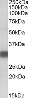
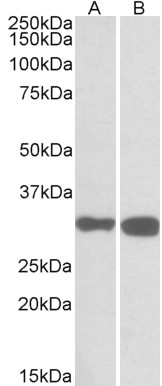
PDXP Antibody (C-Term)
Peptide-affinity purified goat antibody
- SPECIFICATION
- CITATIONS
- PROTOCOLS
- BACKGROUND

Application
| WB, E |
|---|---|
| Primary Accession | Q96GD0 |
| Other Accession | NP_064711.1, 57026, 57028 (mouse), 727679 (rat) |
| Reactivity | Human, Mouse, Rat |
| Predicted | Cow |
| Host | Goat |
| Clonality | Polyclonal |
| Concentration | 0.5 mg/ml |
| Isotype | IgG |
| Calculated MW | 31698 Da |
| Gene ID | 57026 |
|---|---|
| Other Names | Pyridoxal phosphate phosphatase, PLP phosphatase, 3.1.3.3, 3.1.3.74, Chronophin, PDXP, CIN, PLP, PLPP |
| Format | 0.5 mg/ml in Tris saline, 0.02% sodium azide, pH7.3 with 0.5% bovine serum albumin |
| Storage | Maintain refrigerated at 2-8°C for up to 6 months. For long term storage store at -20°C in small aliquots to prevent freeze-thaw cycles. |
| Precautions | PDXP Antibody (C-Term) is for research use only and not for use in diagnostic or therapeutic procedures. |
| Name | PDXP (HGNC:30259) |
|---|---|
| Function | Functions as a pyridoxal phosphate (PLP) phosphatase, which also catalyzes the dephosphorylation of pyridoxine 5'-phosphate (PNP) and pyridoxamine 5'-phosphate (PMP), with order of substrate preference PLP > PNP > PMP and therefore plays a role in vitamin B6 metabolism (PubMed:14522954, PubMed:8132548). Also functions as a protein serine phosphatase that specifically dephosphorylates 'Ser-3' in proteins of the actin-depolymerizing factor (ADF)/cofilin family like CFL1 and DSTN. Thereby, regulates cofilin-dependent actin cytoskeleton reorganization, being required for normal progress through mitosis and normal cytokinesis. Does not dephosphorylate phosphothreonines in LIMK1. Does not dephosphorylate peptides containing phosphotyrosine (PubMed:15580268). |
| Cellular Location | Cytoplasm, cytosol. Cytoplasm, cytoskeleton. Cell projection, ruffle membrane; Peripheral membrane protein; Cytoplasmic side. Cell projection, lamellipodium membrane; Peripheral membrane protein; Cytoplasmic side. Cell membrane; Peripheral membrane protein; Cytoplasmic side. Note=Colocalizes with the actin cytoskeleton in membrane ruffles and lamellipodia. Diffusely distributed throughout the cytosol during pro-metaphase and metaphase Detected at the dynamic cell poles during telophase. Detected at the cleavage furrow and contractile ring during cytokinesis. Transiently detected at the plasma membrane in late stages of cytokinesis. Detected at the midbody. |
| Tissue Location | Ubiquitously expressed (at protein level) (PubMed:23223568). Highly expressed in all the regions of central nerve system except the spinal cord. Also expressed at high level in liver and testis. In fetus, it is weakly expressed in all organs except brain (PubMed:14522954, PubMed:15580268). |

Thousands of laboratories across the world have published research that depended on the performance of antibodies from Abcepta to advance their research. Check out links to articles that cite our products in major peer-reviewed journals, organized by research category.
info@abcepta.com, and receive a free "I Love Antibodies" mug.

Provided below are standard protocols that you may find useful for product applications.
References
Chronophin mediates an ATP-sensing mechanism for cofilin dephosphorylation and neuronal cofilin-actin rod formation. Huang TY, Minamide LS, Bamburg JR, Bokoch GM, Developmental cell 2008 Nov 15 (5): 691-703. PMID: 19000834
If you have used an Abcepta product and would like to share how it has performed, please click on the "Submit Review" button and provide the requested information. Our staff will examine and post your review and contact you if needed.
If you have any additional inquiries please email technical services at tech@abcepta.com.
Ordering Information
Other Products
Shipping Information
