


 Foundational characteristics of cancer include proliferation, angiogenesis, migration, evasion of apoptosis, and cellular immortality. Find key markers for these cellular processes and antibodies to detect them.
 The SUMOplot™ Analysis Program predicts and scores sumoylation sites in your protein. SUMOylation is a post-translational modification involved in various cellular processes, such as nuclear-cytosolic transport, transcriptional regulation, apoptosis, protein stability, response to stress, and progression through the cell cycle.
 The Autophagy Receptor Motif Plotter predicts and scores autophagy receptor binding sites in your protein. Identifying proteins connected to this pathway is critical to understanding the role of autophagy in physiological as well as pathological processes such as development, differentiation, neurodegenerative diseases, stress, infection, and cancer.
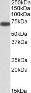
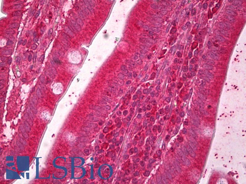
ERO1-like (aa105-118) Antibody (internal region)
Peptide-affinity purified goat antibody

- SPECIFICATION
- CITATIONS
- PROTOCOLS
- BACKGROUND

Application
| WB, IHC, Pep-ELISA |
|---|---|
| Primary Accession | Q96HE7 |
| Other Accession | NP_055399.1, 30001 |
| Reactivity | Human |
| Predicted | Mouse, Rat, Pig, Dog |
| Host | Goat |
| Clonality | Polyclonal |
| Concentration | 0.5 mg/ml |
| Isotype | IgG |
| Calculated MW | 54393 Da |
| Gene ID | 30001 |
|---|---|
| Other Names | ERO1-like protein alpha, ERO1-L, ERO1-L-alpha, 1.8.4.-, Endoplasmic oxidoreductin-1-like protein, Oxidoreductin-1-L-alpha, ERO1L |
| Dilution | WB~~1:1000 IHC~~1:100~500 Pep-ELISA~~N/A |
| Format | 0.5 mg/ml in Tris saline, 0.02% sodium azide, pH7.3 with 0.5% bovine serum albumin |
| Storage | Maintain refrigerated at 2-8°C for up to 6 months. For long term storage store at -20°C in small aliquots to prevent freeze-thaw cycles. |
| Precautions | ERO1-like (aa105-118) Antibody (internal region) is for research use only and not for use in diagnostic or therapeutic procedures. |
| Name | ERO1A (HGNC:13280) |
|---|---|
| Synonyms | ERO1L |
| Function | Oxidoreductase involved in disulfide bond formation in the endoplasmic reticulum. Efficiently reoxidizes P4HB/PDI, the enzyme catalyzing protein disulfide formation, in order to allow P4HB to sustain additional rounds of disulfide formation. Following P4HB reoxidation, passes its electrons to molecular oxygen via FAD, leading to the production of reactive oxygen species (ROS) in the cell. Required for the proper folding of immunoglobulins (PubMed:29858230). Plays an important role in ER stress-induced, CHOP-dependent apoptosis by activating the inositol 1,4,5-trisphosphate receptor IP3R1. Involved in the release of the unfolded cholera toxin from reduced P4HB/PDI in case of infection by V.cholerae, thereby playing a role in retrotranslocation of the toxin. |
| Cellular Location | Endoplasmic reticulum membrane; Peripheral membrane protein; Lumenal side. Golgi apparatus lumen. Secreted. Cell projection, dendrite {ECO:0000250|UniProtKB:Q8R4A1}. Note=The association with ERP44 is essential for its retention in the endoplasmic reticulum (PubMed:29858230). In neurons, it localizes to dendrites (By similarity). {ECO:0000250|UniProtKB:Q8R4A1, ECO:0000269|PubMed:29858230} |
| Tissue Location | Widely expressed at low level. Expressed at high level in upper digestive tract. Highly expressed in esophagus. Weakly expressed in stomach and duodenum. |

Thousands of laboratories across the world have published research that depended on the performance of antibodies from Abcepta to advance their research. Check out links to articles that cite our products in major peer-reviewed journals, organized by research category.
info@abcepta.com, and receive a free "I Love Antibodies" mug.

Provided below are standard protocols that you may find useful for product applications.
References
Crystal structures of human Ero1? reveal the mechanisms of regulated and targeted oxidation of PDI. Inaba K, Masui S, Iida H, Vavassori S, Sitia R, Suzuki M. EMBO J. 2010 Oct 6;29(19):3330-43. PMID: 20834232
If you have used an Abcepta product and would like to share how it has performed, please click on the "Submit Review" button and provide the requested information. Our staff will examine and post your review and contact you if needed.
If you have any additional inquiries please email technical services at tech@abcepta.com.
Ordering Information
Other Products
Shipping Information
