


 Foundational characteristics of cancer include proliferation, angiogenesis, migration, evasion of apoptosis, and cellular immortality. Find key markers for these cellular processes and antibodies to detect them.
 The SUMOplot™ Analysis Program predicts and scores sumoylation sites in your protein. SUMOylation is a post-translational modification involved in various cellular processes, such as nuclear-cytosolic transport, transcriptional regulation, apoptosis, protein stability, response to stress, and progression through the cell cycle.
 The Autophagy Receptor Motif Plotter predicts and scores autophagy receptor binding sites in your protein. Identifying proteins connected to this pathway is critical to understanding the role of autophagy in physiological as well as pathological processes such as development, differentiation, neurodegenerative diseases, stress, infection, and cancer.
> home > Products > Primary Antibodies > Antibody Collections > Goat Antibodies > JAG1 Antibody (C-Term)
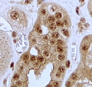
JAG1 Antibody (C-Term)
Peptide-affinity purified goat antibody
- SPECIFICATION
- CITATIONS: 1
- PROTOCOLS
- BACKGROUND

Application
| IHC, E |
|---|---|
| Primary Accession | P78504 |
| Other Accession | NP_000205.1, 182, 16449 (mouse), 29146 (rat) |
| Reactivity | Human |
| Predicted | Mouse, Rat, Pig, Dog |
| Host | Goat |
| Clonality | Polyclonal |
| Concentration | 0.5 mg/ml |
| Isotype | IgG |
| Calculated MW | 133799 Da |
| Gene ID | 182 |
|---|---|
| Other Names | Protein jagged-1, Jagged1, hJ1, CD339, JAG1, JAGL1 |
| Dilution | IHC~~1:100~500 E~~N/A |
| Format | 0.5 mg/ml in Tris saline, 0.02% sodium azide, pH7.3 with 0.5% bovine serum albumin |
| Storage | Maintain refrigerated at 2-8°C for up to 6 months. For long term storage store at -20°C in small aliquots to prevent freeze-thaw cycles. |
| Precautions | JAG1 Antibody (C-Term) is for research use only and not for use in diagnostic or therapeutic procedures. |
| Name | JAG1 |
|---|---|
| Synonyms | JAGL1 |
| Function | Ligand for multiple Notch receptors and involved in the mediation of Notch signaling (PubMed:18660822, PubMed:20437614). May be involved in cell-fate decisions during hematopoiesis (PubMed:9462510). Seems to be involved in early and late stages of mammalian cardiovascular development. Inhibits myoblast differentiation (By similarity). Enhances fibroblast growth factor-induced angiogenesis (in vitro). |
| Cellular Location | Membrane; Single-pass type I membrane protein. Cell membrane |
| Tissue Location | Widely expressed in adult and fetal tissues. In cervix epithelium expressed in undifferentiated subcolumnar reserve cells and squamous metaplasia. Expression is up-regulated in cervical squamous cell carcinoma. Expressed in bone marrow cell line HS-27a which supports the long-term maintenance of immature progenitor cells |
Research Areas
Citations ( 0 )

Application Protocols
Provided below are standard protocols that you may find useful for product applications.
References
Plasminogen activator uPA is a direct transcriptional target of the JAG1-Notch receptor signaling pathway in breast cancer. Shimizu M, Cohen B, Goldvasser P, Berman H, Virtanen C, Reedijk M. Cancer Res. 2011 Jan 1;71(1):277-86. PMID: 21199807
Abcepta welcomes feedback from its customers.
If you have used an Abcepta product and would like to share how it has performed, please click on the "Submit Review" button and provide the requested information. Our staff will examine and post your review and contact you if needed.
If you have any additional inquiries please email technical services at tech@abcepta.com.
$ 423.00
Cat# AF3557a
Ordering Information
United States
AlbaniaAustraliaAustriaBelgiumBosnia & HerzegovinaBrazilBulgariaCanadaCentral AmericaChinaCroatiaCyprusCzech RepublicDenmarkEstoniaFinlandFranceGermanyGreeceHong KongHungaryIcelandIndiaIndonesiaIrelandIsraelItalyJapanLatviaLithuaniaLuxembourgMacedoniaMalaysiaMaltaMexicoNetherlandsNew ZealandNorwayPakistanPolandPortugalRomaniaSerbiaSingaporeSlovakiaSloveniaSouth AfricaSouth KoreaSpainSwedenSwitzerlandTaiwanTurkeyUnited KingdomUnited StatesVietnamWorldwideOthers
USA Headquarters
(888) 735-7227 / (858) 622-0099 or (858) 875-1900
Other Products
Shipping Information
Domestic orders (in stock items)
Shipped out the same day. Orders placed after 1 PM (PST) will ship out the next business day.
International orders
Contact your local distributors
