


 Foundational characteristics of cancer include proliferation, angiogenesis, migration, evasion of apoptosis, and cellular immortality. Find key markers for these cellular processes and antibodies to detect them.
 The SUMOplot™ Analysis Program predicts and scores sumoylation sites in your protein. SUMOylation is a post-translational modification involved in various cellular processes, such as nuclear-cytosolic transport, transcriptional regulation, apoptosis, protein stability, response to stress, and progression through the cell cycle.
 The Autophagy Receptor Motif Plotter predicts and scores autophagy receptor binding sites in your protein. Identifying proteins connected to this pathway is critical to understanding the role of autophagy in physiological as well as pathological processes such as development, differentiation, neurodegenerative diseases, stress, infection, and cancer.
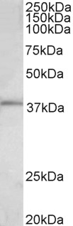
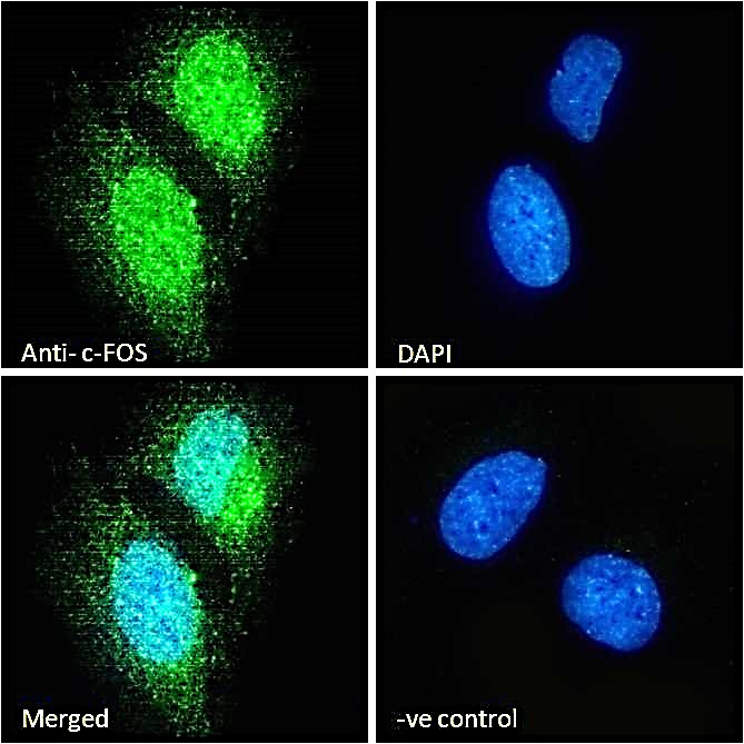
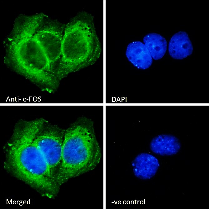
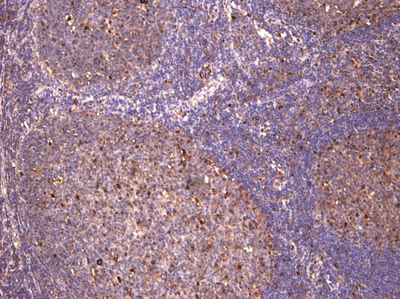
c-FOS (aa283-295) Antibody (internal region)
Peptide-affinity purified goat antibody

- SPECIFICATION
- CITATIONS
- PROTOCOLS
- BACKGROUND

Application
| IHC, IF, Pep-ELISA |
|---|---|
| Primary Accession | P01100 |
| Other Accession | NP_005243.1, 2353 |
| Reactivity | Human |
| Predicted | Pig, Dog, Cat |
| Host | Goat |
| Clonality | Polyclonal |
| Concentration | 0.5 mg/ml |
| Isotype | IgG |
| Calculated MW | 40695 Da |
| Gene ID | 2353 |
|---|---|
| Other Names | Proto-oncogene c-Fos, Cellular oncogene fos, G0/G1 switch regulatory protein 7, FOS, G0S7 |
| Dilution | IHC~~1:100~500 IF~~1:50~200 Pep-ELISA~~N/A |
| Format | 0.5 mg/ml in Tris saline, 0.02% sodium azide, pH7.3 with 0.5% bovine serum albumin |
| Storage | Maintain refrigerated at 2-8°C for up to 6 months. For long term storage store at -20°C in small aliquots to prevent freeze-thaw cycles. |
| Precautions | c-FOS (aa283-295) Antibody (internal region) is for research use only and not for use in diagnostic or therapeutic procedures. |
| Name | FOS |
|---|---|
| Synonyms | G0S7 |
| Function | Nuclear phosphoprotein which forms a tight but non-covalently linked complex with the JUN/AP-1 transcription factor. In the heterodimer, FOS and JUN/AP-1 basic regions each seems to interact with symmetrical DNA half sites. On TGF-beta activation, forms a multimeric SMAD3/SMAD4/JUN/FOS complex at the AP1/SMAD-binding site to regulate TGF-beta-mediated signaling. Has a critical function in regulating the development of cells destined to form and maintain the skeleton. It is thought to have an important role in signal transduction, cell proliferation and differentiation. In growing cells, activates phospholipid synthesis, possibly by activating CDS1 and PI4K2A. This activity requires Tyr-dephosphorylation and association with the endoplasmic reticulum. |
| Cellular Location | Nucleus. Endoplasmic reticulum. Cytoplasm, cytosol. Note=In quiescent cells, present in very small amounts in the cytosol. Following induction of cell growth, first localizes to the endoplasmic reticulum and only later to the nucleus. Localization at the endoplasmic reticulum requires dephosphorylation at Tyr-10 and Tyr- 30 |

Thousands of laboratories across the world have published research that depended on the performance of antibodies from Abcepta to advance their research. Check out links to articles that cite our products in major peer-reviewed journals, organized by research category.
info@abcepta.com, and receive a free "I Love Antibodies" mug.

Provided below are standard protocols that you may find useful for product applications.
References
c-Fos regulates hepatitis C virus propagation. Kang SM, Lim S, Won SJ, Shin YJ, Lim YS, Ahn BY, Hwang SB. FEBS Lett. 2011 Oct 20;585(20):3236-44. PMID: 21920361
If you have used an Abcepta product and would like to share how it has performed, please click on the "Submit Review" button and provide the requested information. Our staff will examine and post your review and contact you if needed.
If you have any additional inquiries please email technical services at tech@abcepta.com.
Ordering Information
Other Products
Shipping Information
