


 Foundational characteristics of cancer include proliferation, angiogenesis, migration, evasion of apoptosis, and cellular immortality. Find key markers for these cellular processes and antibodies to detect them.
 The SUMOplot™ Analysis Program predicts and scores sumoylation sites in your protein. SUMOylation is a post-translational modification involved in various cellular processes, such as nuclear-cytosolic transport, transcriptional regulation, apoptosis, protein stability, response to stress, and progression through the cell cycle.
 The Autophagy Receptor Motif Plotter predicts and scores autophagy receptor binding sites in your protein. Identifying proteins connected to this pathway is critical to understanding the role of autophagy in physiological as well as pathological processes such as development, differentiation, neurodegenerative diseases, stress, infection, and cancer.
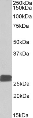
DUSP6 / MKP3 Antibody (C-Term)
Peptide-affinity purified goat antibody
- SPECIFICATION
- CITATIONS
- PROTOCOLS
- BACKGROUND

Application
| WB, E |
|---|---|
| Primary Accession | Q16828 |
| Other Accession | NP_001937.2, NP_073143.2, 1848, 67603 (mouse), 116663 (rat) |
| Reactivity | Human, Mouse, Rat, Pig |
| Predicted | Dog |
| Host | Goat |
| Clonality | Polyclonal |
| Concentration | 0.5 mg/ml |
| Isotype | IgG |
| Calculated MW | 42320 Da |
| Gene ID | 1848 |
|---|---|
| Other Names | Dual specificity protein phosphatase 6, 3.1.3.16, 3.1.3.48, Dual specificity protein phosphatase PYST1, Mitogen-activated protein kinase phosphatase 3, MAP kinase phosphatase 3, MKP-3, DUSP6, MKP3, PYST1 |
| Dilution | WB~~1:1000 E~~N/A |
| Format | 0.5 mg/ml in Tris saline, 0.02% sodium azide, pH7.3 with 0.5% bovine serum albumin |
| Storage | Maintain refrigerated at 2-8°C for up to 6 months. For long term storage store at -20°C in small aliquots to prevent freeze-thaw cycles. |
| Precautions | DUSP6 / MKP3 Antibody (C-Term) is for research use only and not for use in diagnostic or therapeutic procedures. |
| Name | DUSP6 |
|---|---|
| Synonyms | MKP3, PYST1 |
| Function | Dual specificity protein phosphatase, which mediates dephosphorylation and inactivation of MAP kinases (PubMed:8670865). Has a specificity for the ERK family (PubMed:8670865). Plays an important role in alleviating chronic postoperative pain (By similarity). Necessary for the normal dephosphorylation of the long-lasting phosphorylated forms of spinal MAPK1/3 and MAP kinase p38 induced by peripheral surgery, which drives the resolution of acute postoperative allodynia (By similarity). Also important for dephosphorylation of MAPK1/3 in local wound tissue, which further contributes to resolution of acute pain (By similarity). Promotes cell differentiation by regulating MAPK1/MAPK3 activity and regulating the expression of AP1 transcription factors (PubMed:29043977). |
| Cellular Location | Cytoplasm. |
| Tissue Location | Expressed in keratinocytes (at protein level). |

Thousands of laboratories across the world have published research that depended on the performance of antibodies from Abcepta to advance their research. Check out links to articles that cite our products in major peer-reviewed journals, organized by research category.
info@abcepta.com, and receive a free "I Love Antibodies" mug.

Provided below are standard protocols that you may find useful for product applications.
Background
This antibody is expected to recognize both reported isoforms (NP_001937.2; NP_073143.2).
References
Signal integration and coincidence detection in the mitogen-activated protein kinase/extracellular signal-regulated kinase (ERK) cascade: concomitant activation of receptor tyrosine kinases and of LRP-1 leads to sustained ERK phosphorylation via down-regu Geetha N, Mihaly J, Stockenhuber A, Blasi F, Uhrin P, Binder BR, Freissmuth M, Breuss JM. J Biol Chem. 2011 Jul 22;286(29):25663-74. PMID: 21610072
If you have used an Abcepta product and would like to share how it has performed, please click on the "Submit Review" button and provide the requested information. Our staff will examine and post your review and contact you if needed.
If you have any additional inquiries please email technical services at tech@abcepta.com.
Ordering Information
Other Products
Shipping Information
