


 Foundational characteristics of cancer include proliferation, angiogenesis, migration, evasion of apoptosis, and cellular immortality. Find key markers for these cellular processes and antibodies to detect them.
 The SUMOplot™ Analysis Program predicts and scores sumoylation sites in your protein. SUMOylation is a post-translational modification involved in various cellular processes, such as nuclear-cytosolic transport, transcriptional regulation, apoptosis, protein stability, response to stress, and progression through the cell cycle.
 The Autophagy Receptor Motif Plotter predicts and scores autophagy receptor binding sites in your protein. Identifying proteins connected to this pathway is critical to understanding the role of autophagy in physiological as well as pathological processes such as development, differentiation, neurodegenerative diseases, stress, infection, and cancer.
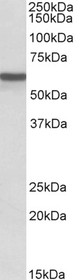
ADRA1B Antibody (internal region)
Peptide-affinity purified goat antibody
- SPECIFICATION
- CITATIONS
- PROTOCOLS
- BACKGROUND

Application
| WB, E |
|---|---|
| Primary Accession | P35368 |
| Other Accession | NP_000670.1, 147, 11548 (mouse), 24173 (rat) |
| Reactivity | Human, Mouse, Rat, Pig |
| Predicted | Dog |
| Host | Goat |
| Clonality | Polyclonal |
| Concentration | 0.5 mg/ml |
| Isotype | IgG |
| Calculated MW | 56836 Da |
| Gene ID | 147 |
|---|---|
| Other Names | Alpha-1B adrenergic receptor, Alpha-1B adrenoreceptor, Alpha-1B adrenoceptor, ADRA1B |
| Dilution | WB~~1:1000 E~~N/A |
| Format | 0.5 mg/ml in Tris saline, 0.02% sodium azide, pH7.3 with 0.5% bovine serum albumin |
| Storage | Maintain refrigerated at 2-8°C for up to 6 months. For long term storage store at -20°C in small aliquots to prevent freeze-thaw cycles. |
| Precautions | ADRA1B Antibody (internal region) is for research use only and not for use in diagnostic or therapeutic procedures. |
| Name | ADRA1B |
|---|---|
| Function | This alpha-adrenergic receptor mediates its action by association with G proteins that activate a phosphatidylinositol- calcium second messenger system. Its effect is mediated by G(q) and G(11) proteins. Nuclear ADRA1A-ADRA1B heterooligomers regulate phenylephrine (PE)-stimulated ERK signaling in cardiac myocytes. |
| Cellular Location | Nucleus membrane; Multi-pass membrane protein. Cell membrane; Multi-pass membrane protein. Cytoplasm Membrane, caveola. Note=Location at the nuclear membrane facilitates heterooligomerization and regulates ERK- mediated signaling in cardiac myocytes. signaling in cardiac myocytes Colocalizes with GNAQ, PLCB1 as well as LAP2 at the nuclear membrane of cardiac myocytes |

Thousands of laboratories across the world have published research that depended on the performance of antibodies from Abcepta to advance their research. Check out links to articles that cite our products in major peer-reviewed journals, organized by research category.
info@abcepta.com, and receive a free "I Love Antibodies" mug.

Provided below are standard protocols that you may find useful for product applications.
References
Alpha- and beta-adrenergic receptor (AR) protein expression is associated with poor clinical outcome in breast cancer: an immunohistochemical study. Powe DG, Voss MJ, Habashy HO, Zänker KS, Green AR, Ellis IO, Entschladen F. Breast Cancer Res Treat. 2011 Nov;130(2):457-63. PMID: 21298476
If you have used an Abcepta product and would like to share how it has performed, please click on the "Submit Review" button and provide the requested information. Our staff will examine and post your review and contact you if needed.
If you have any additional inquiries please email technical services at tech@abcepta.com.
Ordering Information
Other Products
Shipping Information
