


 Foundational characteristics of cancer include proliferation, angiogenesis, migration, evasion of apoptosis, and cellular immortality. Find key markers for these cellular processes and antibodies to detect them.
 The SUMOplot™ Analysis Program predicts and scores sumoylation sites in your protein. SUMOylation is a post-translational modification involved in various cellular processes, such as nuclear-cytosolic transport, transcriptional regulation, apoptosis, protein stability, response to stress, and progression through the cell cycle.
 The Autophagy Receptor Motif Plotter predicts and scores autophagy receptor binding sites in your protein. Identifying proteins connected to this pathway is critical to understanding the role of autophagy in physiological as well as pathological processes such as development, differentiation, neurodegenerative diseases, stress, infection, and cancer.
Goat Anti-SERPINF1 / PEDF Antibody (internal region)
Purified Goat Polyclonal Antibody
- SPECIFICATION
- CITATIONS
- PROTOCOLS
- BACKGROUND

Application
| WB, E |
|---|---|
| Primary Accession | P36955 |
| Other Accession | NP_002606.3 |
| Reactivity | Human |
| Predicted | Human, Pig, Dog |
| Host | Goat |
| Clonality | Polyclonal |
| Concentration | 0.5 |
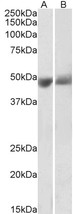
| Calculated MW | 46312 Da |
| Gene ID | 5176 |
|---|---|
| Other Names | SERPINF1; EPC-1; PEDF; PIG35; serpin peptidase inhibitor, clade F (alpha-2 antiplasmin, pigment epithelium derived factor), member 1; pigment epithelium-derived factor; proliferation-inducing protein 35; serine (or cysteine) proteinase inhibitor, clade F (alpha-2 antiplasmin, pigment epithelium derived factor), member 1 |
| Dilution | WB~~1:1000 E~~N/A |
| Format | Supplied at 0.5 mg/ml in Tris saline, 0.02% sodium azide, pH7.3 with 0.5% bovine serum albumin. Aliquot and store at -20°C. Minimize freezing and thawing. |
| Immunogen | Peptide with sequence C-KITGKPIKLTQVEHR, from the internal region of the protein sequence according to NP_002606.3. |
| Storage | Maintain refrigerated at 2-8°C for up to 6 months. For long term storage store at -20°C in small aliquots to prevent freeze-thaw cycles. |
| Precautions | Goat Anti-SERPINF1 / PEDF Antibody (internal region) is for research use only and not for use in diagnostic or therapeutic procedures. |
| Name | SERPINF1 |
|---|---|
| Synonyms | PEDF |
| Function | Neurotrophic protein; induces extensive neuronal differentiation in retinoblastoma cells. Potent inhibitor of angiogenesis. As it does not undergo the S (stressed) to R (relaxed) conformational transition characteristic of active serpins, it exhibits no serine protease inhibitory activity. |
| Cellular Location | Secreted. Melanosome. Note=Enriched in stage I melanosomes |
| Tissue Location | Retinal pigment epithelial cells and blood plasma. |

Thousands of laboratories across the world have published research that depended on the performance of antibodies from Abcepta to advance their research. Check out links to articles that cite our products in major peer-reviewed journals, organized by research category.
info@abcepta.com, and receive a free "I Love Antibodies" mug.

Provided below are standard protocols that you may find useful for product applications.
References
pposing effects of pigment epithelium-derived factor on breast cancer cell versus neuronal survival: implication for brain metastasis and metastasis-induced brain damage. Fitzgerald DP et al. Cancer Res. 2012 Jan 1;72(1):144-53.
If you have used an Abcepta product and would like to share how it has performed, please click on the "Submit Review" button and provide the requested information. Our staff will examine and post your review and contact you if needed.
If you have any additional inquiries please email technical services at tech@abcepta.com.
Ordering Information
Other Products
Shipping Information
