


 Foundational characteristics of cancer include proliferation, angiogenesis, migration, evasion of apoptosis, and cellular immortality. Find key markers for these cellular processes and antibodies to detect them.
 The SUMOplot™ Analysis Program predicts and scores sumoylation sites in your protein. SUMOylation is a post-translational modification involved in various cellular processes, such as nuclear-cytosolic transport, transcriptional regulation, apoptosis, protein stability, response to stress, and progression through the cell cycle.
 The Autophagy Receptor Motif Plotter predicts and scores autophagy receptor binding sites in your protein. Identifying proteins connected to this pathway is critical to understanding the role of autophagy in physiological as well as pathological processes such as development, differentiation, neurodegenerative diseases, stress, infection, and cancer.
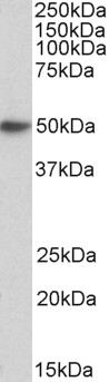
Goat Anti-FCER1A Antibody (internal region)
Purified Goat Polyclonal Antibody
- SPECIFICATION
- CITATIONS
- PROTOCOLS
- BACKGROUND

Application
| WB, E |
|---|---|
| Primary Accession | P12319 |
| Other Accession | NP_001992.1 |
| Reactivity | Human |
| Predicted | Human |
| Host | Goat |
| Clonality | Polyclonal |
| Concentration | 0.5 |
| Calculated MW | 29596 Da |
| Gene ID | 2205 |
|---|---|
| Other Names | FCER1A; Fc fragment of IgE, high affinity I, receptor for; alpha polypeptide; FCE1A; FcERI; Fc IgE receptor, alpha polypeptide; Fc epsilon RI alpha-chain; Fc-epsilon RI-alpha; high affinity immunoglobulin epsilon receptor alpha-subunit; high affinity immunoglobulin epsilon receptor subunit alpha; igE Fc receptor subunit alpha; immunoglobulin E receptor, high-affinity, of mast cells, alpha polypeptide |
| Dilution | WB~~1:1000 E~~N/A |
| Format | Supplied at 0.5 mg/ml in Tris saline, 0.02% sodium azide, pH7.3 with 0.5% bovine serum albumin. Aliquot and store at -20°C. Minimize freezing and thawing. |
| Immunogen | Peptide with sequence C-TGKVWQLDYESEP, from the internal region of the protein sequence according to NP_001992.1. |
| Storage | Maintain refrigerated at 2-8°C for up to 6 months. For long term storage store at -20°C in small aliquots to prevent freeze-thaw cycles. |
| Precautions | Goat Anti-FCER1A Antibody (internal region) is for research use only and not for use in diagnostic or therapeutic procedures. |
| Name | FCER1A |
|---|---|
| Synonyms | FCE1A |
| Function | High-affinity receptor for immunoglobulin epsilon/IgE. Mediates IgE effector functions in myeloid cells. Upon IgE binding and antigen/allergen cross-linking initiates signaling pathways that lead to myeloid cell activation and differentiation. On mast cells, basophils and eosinophils stimulates the secretion of vasoactive amines, lipid mediators and cytokines that contribute to inflammatory response, tissue remodeling and cytotoxicity against microbes. Triggers the immediate hypersensitivity response to allergens as a host defense mechanism against helminth parasites, pathogenic bacteria and venom toxicity. When dysregulated, it can elicit harmful life-threatening allergic and anaphylactic reactions. |
| Cellular Location | Cell membrane; Single-pass type I membrane protein |
| Tissue Location | Expressed in eosinophils. |

Thousands of laboratories across the world have published research that depended on the performance of antibodies from Abcepta to advance their research. Check out links to articles that cite our products in major peer-reviewed journals, organized by research category.
info@abcepta.com, and receive a free "I Love Antibodies" mug.

Provided below are standard protocols that you may find useful for product applications.
References
Expression of high-affinity IgE receptor on human peripheral blood dendritic cells in children. Vasudev M, Cheung DS, Pincsak H, Li SH, Yan K, Simpson P, Dasu T, Grayson MH. PloS one 2012 7 (2): e32556.
If you have used an Abcepta product and would like to share how it has performed, please click on the "Submit Review" button and provide the requested information. Our staff will examine and post your review and contact you if needed.
If you have any additional inquiries please email technical services at tech@abcepta.com.

Ordering Information
Other Products
Shipping Information
