


 Foundational characteristics of cancer include proliferation, angiogenesis, migration, evasion of apoptosis, and cellular immortality. Find key markers for these cellular processes and antibodies to detect them.
 The SUMOplot™ Analysis Program predicts and scores sumoylation sites in your protein. SUMOylation is a post-translational modification involved in various cellular processes, such as nuclear-cytosolic transport, transcriptional regulation, apoptosis, protein stability, response to stress, and progression through the cell cycle.
 The Autophagy Receptor Motif Plotter predicts and scores autophagy receptor binding sites in your protein. Identifying proteins connected to this pathway is critical to understanding the role of autophagy in physiological as well as pathological processes such as development, differentiation, neurodegenerative diseases, stress, infection, and cancer.
Goat Anti-HLA-B Antibody (internal region)
Purified Goat Polyclonal Antibody
- SPECIFICATION
- CITATIONS
- PROTOCOLS
- BACKGROUND

Application
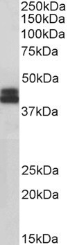
| WB, E |
|---|---|
| Primary Accession | P01889 |
| Other Accession | NP_005505.2 |
| Reactivity | Human |
| Predicted | Human |
| Host | Goat |
| Clonality | Polyclonal |
| Concentration | 0.5 |
| Calculated MW | 40460 Da |
| Gene ID | 3106 |
|---|---|
| Other Names | HLA-B; major histocompatibility complex, class I, B; AS; HLAB; SPDA1; HLA class I histocompatibility antigen, B alpha chain; MHC Class I HLA heavy chain; MHC HLA-B cell surface glycoprotein; MHC HLA-B transmembrane glycoprotein; MHC class I antigen GN0010 |
| Dilution | WB~~1:1000 E~~N/A |
| Format | Supplied at 0.5 mg/ml in Tris saline, 0.02% sodium azide, pH7.3 with 0.5% bovine serum albumin. Aliquot and store at -20°C. Minimize freezing and thawing. |
| Immunogen | Peptide with sequence C-RLLRGHDQYAYD , from the internal region of the protein sequence according to NP_005505.2. |
| Storage | Maintain refrigerated at 2-8°C for up to 6 months. For long term storage store at -20°C in small aliquots to prevent freeze-thaw cycles. |
| Precautions | Goat Anti-HLA-B Antibody (internal region) is for research use only and not for use in diagnostic or therapeutic procedures. |
| Name | HLA-B (HGNC:4932) |
|---|---|
| Synonyms | HLAB |
| Function | Antigen-presenting major histocompatibility complex class I (MHCI) molecule. In complex with B2M/beta 2 microglobulin displays primarily viral and tumor-derived peptides on antigen-presenting cells for recognition by alpha-beta T cell receptor (TCR) on HLA-B-restricted CD8-positive T cells, guiding antigen-specific T cell immune response to eliminate infected or transformed cells (PubMed:23209413, PubMed:25808313, PubMed:29531227, PubMed:9620674). May also present self-peptides derived from the signal sequence of secreted or membrane proteins, although T cells specific for these peptides are usually inactivated to prevent autoreactivity (PubMed:18991276, PubMed:7743181). Both the peptide and the MHC molecule are recognized by TCR, the peptide is responsible for the fine specificity of antigen recognition and MHC residues account for the MHC restriction of T cells (PubMed:24600035, PubMed:29531227, PubMed:9620674). Typically presents intracellular peptide antigens of 8 to 13 amino acids that arise from cytosolic proteolysis via constitutive proteasome and IFNG-induced immunoproteasome (PubMed:23209413). Can bind different peptides containing allele-specific binding motifs, which are mainly defined by anchor residues at position 2 and 9 (PubMed:25808313, PubMed:29531227). |
| Cellular Location | Cell membrane; Single-pass type I membrane protein. Endoplasmic reticulum membrane; Single-pass type I membrane protein |

Thousands of laboratories across the world have published research that depended on the performance of antibodies from Abcepta to advance their research. Check out links to articles that cite our products in major peer-reviewed journals, organized by research category.
info@abcepta.com, and receive a free "I Love Antibodies" mug.

Provided below are standard protocols that you may find useful for product applications.
References
Biosynthesis of HLA-A and HLA-B antigens in vivo. Owen MJ, Kissonerghis AM, Lodish HF. The Journal of biological chemistry 1980 Oct 255 (20): 9678-84.
If you have used an Abcepta product and would like to share how it has performed, please click on the "Submit Review" button and provide the requested information. Our staff will examine and post your review and contact you if needed.
If you have any additional inquiries please email technical services at tech@abcepta.com.
Ordering Information
Other Products
Shipping Information
