


 Foundational characteristics of cancer include proliferation, angiogenesis, migration, evasion of apoptosis, and cellular immortality. Find key markers for these cellular processes and antibodies to detect them.
 The SUMOplot™ Analysis Program predicts and scores sumoylation sites in your protein. SUMOylation is a post-translational modification involved in various cellular processes, such as nuclear-cytosolic transport, transcriptional regulation, apoptosis, protein stability, response to stress, and progression through the cell cycle.
 The Autophagy Receptor Motif Plotter predicts and scores autophagy receptor binding sites in your protein. Identifying proteins connected to this pathway is critical to understanding the role of autophagy in physiological as well as pathological processes such as development, differentiation, neurodegenerative diseases, stress, infection, and cancer.
> home > Products > Primary Antibodies > Antibody Collections > Skin Melanoma positive > KD-Validated Anti-MHC class 1 Rabbit Monoclonal Antibody
KD-Validated Anti-MHC class 1 Rabbit Monoclonal Antibody
Rabbit monoclonal antibody
- SPECIFICATION
- CITATIONS
- PROTOCOLS
- BACKGROUND

Application
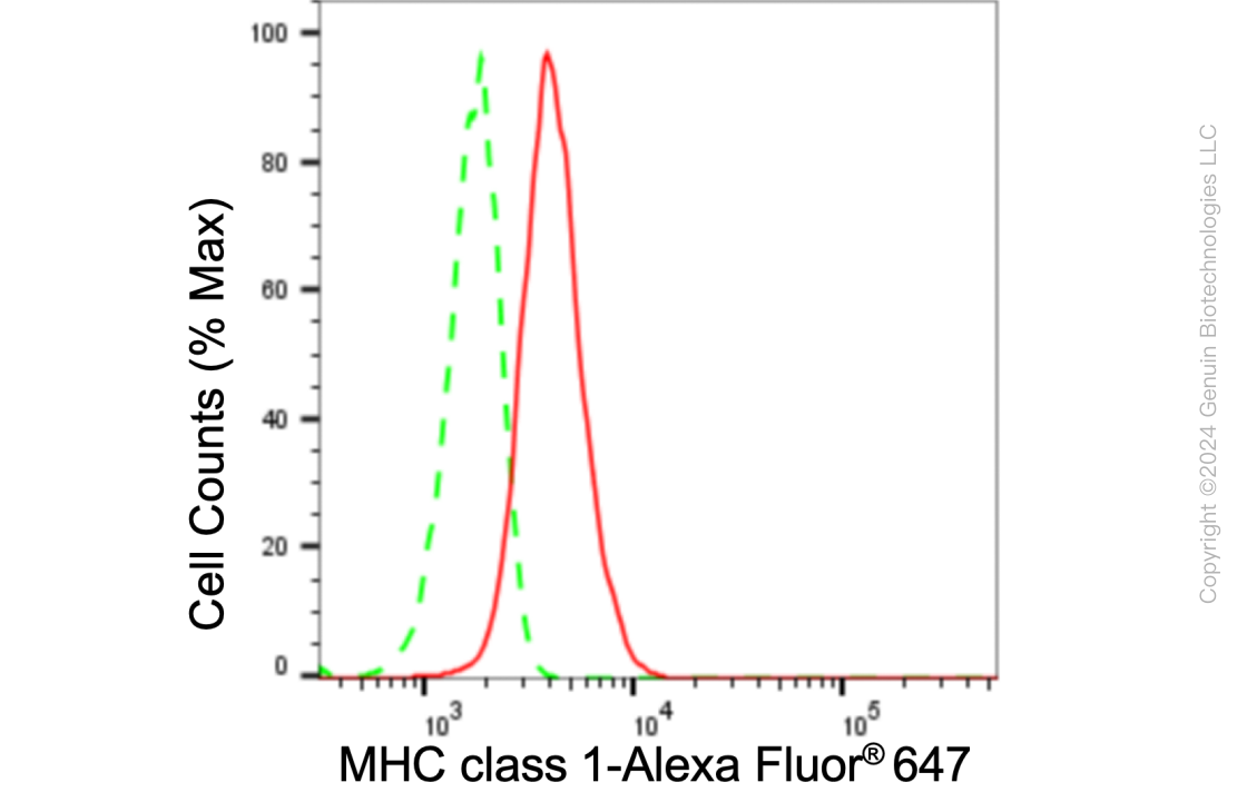
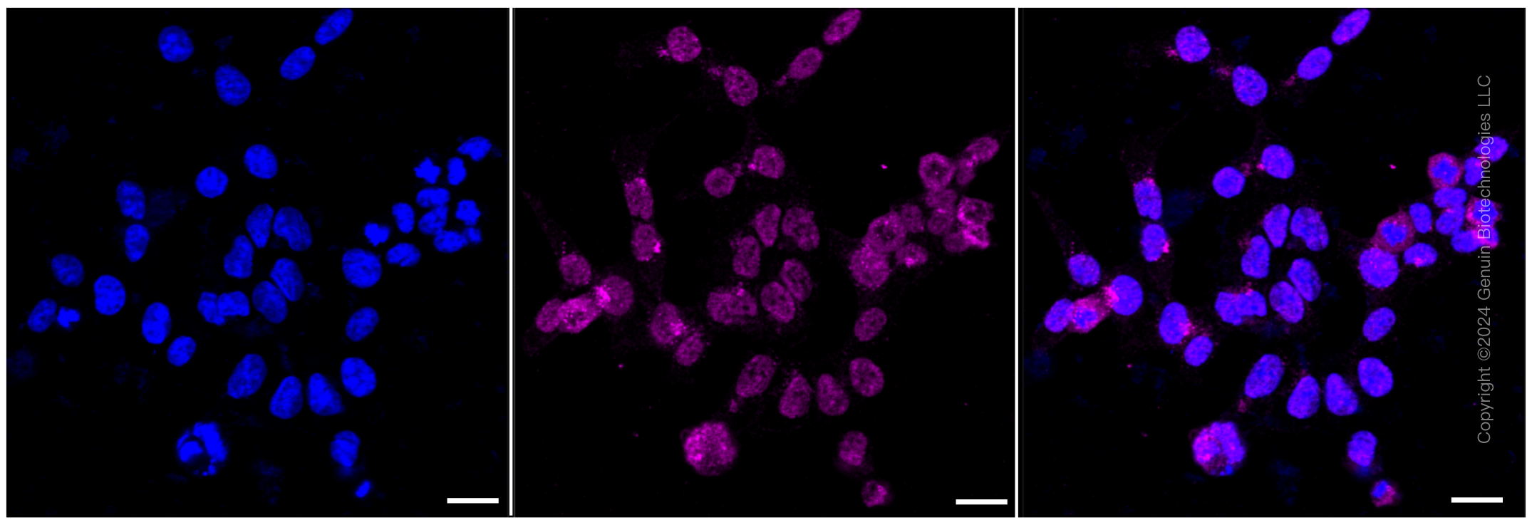
| WB, FC, ICC |
|---|---|
| Primary Accession | P04439 |
| Reactivity | Rat, Human, Mouse |
| Clonality | Monoclonal |
| Isotype | Rabbit IgG |
| Clone Names | 24GB6040 |
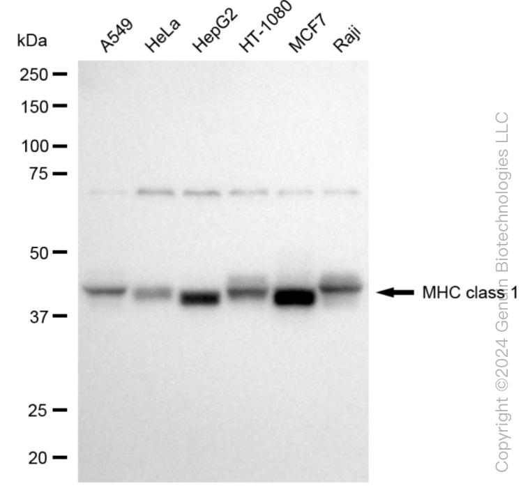
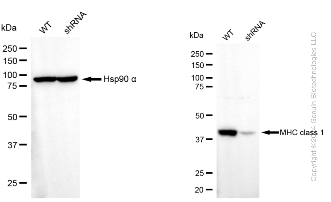
| Calculated MW | Predicted, 41 kDa, observed, 41 kDa |
| Gene Name | HLA-A |
| Aliases | HLA-A; Major Histocompatibility Complex, Class I, A; HLA Class I Histocompatibility Antigen, A Alpha Chain; HLAA; HLA Class I Histocompatibility Antigen, A-1 Alpha Chain; MHC Class I Antigen HLA-A Heavy Chain; Leukocyte Antigen Class I-A; Human Leukocyte Antigen A |
| Immunogen | A synthesized peptide derived from human MHC class 1 |
| Gene ID | 3105 |
|---|---|
| Other Names | HLA class I histocompatibility antigen, A alpha chain, Human leukocyte antigen A, HLA-A, HLA-A (HGNC:4931), HLAA |
| Name | HLA-A (HGNC:4931) |
|---|---|
| Synonyms | HLAA |
| Function | Antigen-presenting major histocompatibility complex class I (MHCI) molecule. In complex with B2M/beta 2 microglobulin displays primarily viral and tumor-derived peptides on antigen-presenting cells for recognition by alpha-beta T cell receptor (TCR) on HLA-A-restricted CD8-positive T cells, guiding antigen-specific T cell immune response to eliminate infected or transformed cells (PubMed:10449296, PubMed:12138174, PubMed:12393434, PubMed:1402688, PubMed:15893615, PubMed:17189421, PubMed:19543285, PubMed:21498667, PubMed:24192765, PubMed:24395804, PubMed:2456340, PubMed:2784196, PubMed:28250417, PubMed:7504010, PubMed:7694806, PubMed:9862734). May also present self- peptides derived from the signal sequence of secreted or membrane proteins, although T cells specific for these peptides are usually inactivated to prevent autoreactivity (PubMed:25880248, PubMed:7506728, PubMed:7679507). Both the peptide and the MHC molecule are recognized by TCR, the peptide is responsible for the fine specificity of antigen recognition and MHC residues account for the MHC restriction of T cells (PubMed:12796775, PubMed:18275829, PubMed:19542454, PubMed:28250417). Typically presents intracellular peptide antigens of 8 to 13 amino acids that arise from cytosolic proteolysis via IFNG-induced immunoproteasome or via endopeptidase IDE/insulin-degrading enzyme (PubMed:17079320, PubMed:17189421, PubMed:20364150, PubMed:26929325, PubMed:27049119). Can bind different peptides containing allele- specific binding motifs, which are mainly defined by anchor residues at position 2 and 9 (PubMed:7504010, PubMed:9862734). |
| Cellular Location | Cell membrane; Single-pass type I membrane protein. Endoplasmic reticulum membrane; Single-pass type I membrane protein |
| Tissue Location | Ubiquitous.. |
Research Areas
Citations (0)

Thousands of laboratories across the world have published research that depended on the performance of antibodies from Abcepta to advance their research. Check out links to articles that cite our products in major peer-reviewed journals, organized by research category.
Submit your citation using an Abcepta antibody to
info@abcepta.com, and receive a free "I Love Antibodies" mug.
info@abcepta.com, and receive a free "I Love Antibodies" mug.

Application Protocols
Provided below are standard protocols that you may find useful for product applications.
Abcepta welcomes feedback from its customers.
If you have used an Abcepta product and would like to share how it has performed, please click on the "Submit Review" button and provide the requested information. Our staff will examine and post your review and contact you if needed.
If you have any additional inquiries please email technical services at tech@abcepta.com.
$ 399.20

$ 149.00
Cat# AGI1048
Ordering Information
United States
AlbaniaAustraliaAustriaBelgiumBosnia & HerzegovinaBrazilBulgariaCanadaCentral AmericaChinaCroatiaCyprusCzech RepublicDenmarkEstoniaFinlandFranceGermanyGreeceHong KongHungaryIcelandIndiaIndonesiaIrelandIsraelItalyJapanLatviaLithuaniaLuxembourgMacedoniaMalaysiaMaltaMexicoNetherlandsNew ZealandNorwayPakistanPolandPortugalRomaniaSerbiaSingaporeSlovakiaSloveniaSouth AfricaSouth KoreaSpainSwedenSwitzerlandTaiwanTurkeyUnited KingdomUnited StatesVietnamWorldwideOthers
USA Headquarters
(888) 735-7227 / (858) 622-0099 or (858) 875-1900
Other Products
Shipping Information
Domestic orders (in stock items)
Shipped out the same day. Orders placed after 1 PM (PST) will ship out the next business day.
International orders
Contact your local distributors
